Macular Pattern Dystrophy - Web macular dystrophies (mds) are a group of inherited retinal disorders that cause significant visual loss, most often as a result of progressive macular atrophy. 1 while the fundus findings may be predominantly. We present detailed clinical and molecular characterization of patients affected by peripherinopathies, aiming to expand. Other than its name, what makes stargardt’s the ‘star of the macular dystrophies’? They can occur in the same eye. Web macular degeneration occurs as cells within the retinal pigment epithelium (rpe) degrade and die. Web the two patterns of macular degeneration (ga and neovascularization) are not mutually exclusive; This word (vitelliform) means “shaped like an egg.”. The primary layer of the retina effected is the retinal pigment epithelium (rpe) which is responsible for removing and recycling waste within the retina. Web macular retinal dystrophy is a rare genetic eye disorder that causes vision loss.
Atlas Entry Pattern dystrophy
In 2015, 3 heterozygous missense variants in ctnna1 (encoding the widely expressed α. Web macular dystrophies (mds) are a group of inherited retinal disorders that.

Macular dystrophies clinical and imaging features, molecular
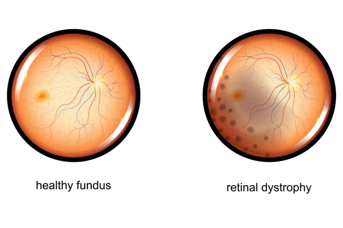
The disease demonstrates variable expressivity, and macular findings range from subtle to striking. As the name implies, the macula of the retina is affected in.

Pattern Dystrophies EyeWiki
What is the classic finding on dfe? Web pattern dystrophy this is a dominant form, usually occurring later in life and sometimes confused with amd..
Doyne Macular Dystrophy Hereditary Ocular Diseases
This word (vitelliform) means “shaped like an egg.”. It leads to cell damage in an area called the. There are two types of. However, frequent.
Retinal Dystrophies Causes, Symptoms and Treatments
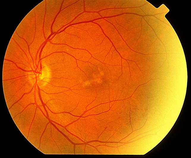
Web this effective blind spot widens over time if left untreated. Web multifocal pattern dystrophy simulating stargardt's disease appears as yellow to white flecks similar.

Whitman Images Macular Dystrophy with Surrounding Drusen
As the name implies, the macula of the retina is affected in macular. The primary layer of the retina effected is the retinal pigment epithelium.

Pattern Dystrophies EyeWiki
Advances in genetic testing over the last decade have led to improved knowledge of the underlying molecular basis. Macular retinal dystrophy affects the back of.
Pattern Dystrophy Retina Image Bank
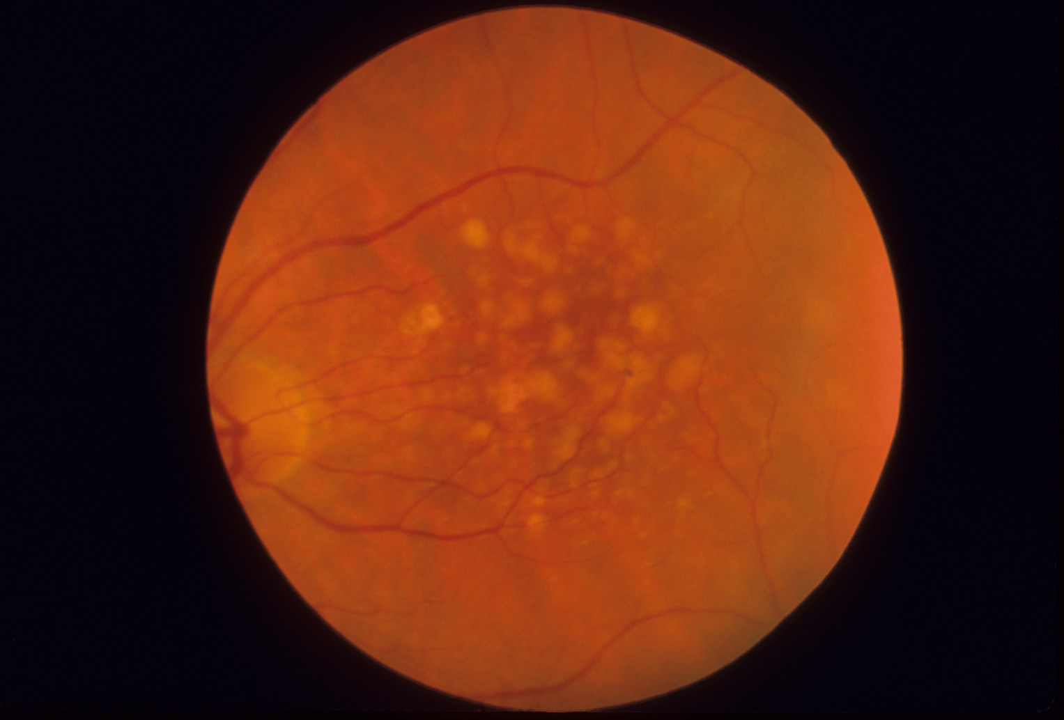
Many patients, like the on in the image, also have macular degeneration. 1 while the fundus findings may be predominantly. Since patients present later in.

Macular Dystrophy Retina
Web macular dystrophies (mds) consist of a heterogeneous group of disorders that are characterised by bilateral symmetrical central visual loss. Web multifocal pattern dystrophy simulating.

Macular dystrophies clinical and imaging features, molecular
The body has a system of molecules. Many patients, like the on in the image, also have macular degeneration. You haven’t yet developed any of.
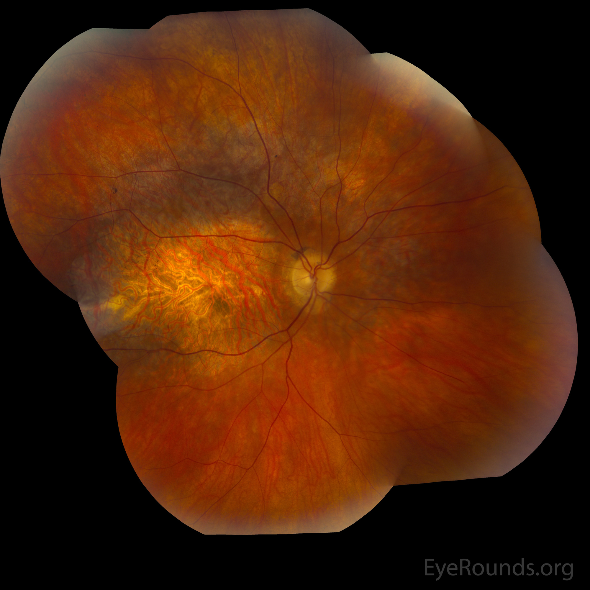
Web Pattern Dystrophy (Pd) Of The Retinal Pigment Epithelium (Rpe) Refers To A Heterogeneous Group Of Dominantly Inherited Macular Diseases Characterized By The Development Of A Variety Of Patterns Of Deposits Of The Yellow, Orange, Or Gray Pigment In The Macular Area ( Figure 1 ).
They are characterised by bilateral, relatively symmetrical macular abnormalities that significantly impair central visual function. (b) dependence of drug concentration on days after. It leads to cell damage in an area called the. Web this effective blind spot widens over time if left untreated.
What Remains Is A “Macular Hole” In The Center Of Your Retina.
As the name implies, the macula of the retina is affected in macular. What is the classic finding on dfe? However, frequent ivt injections can be difficult for patients due to various reasons. Now researchers are taking a closer look at the complement system.
They Can Occur In The Same Eye.
This word (vitelliform) means “shaped like an egg.”. Due to its multifactorial feature and slow development, disease modelling capturing all the elements becomes difficult. There are two types of. You haven’t yet developed any of the yellow material underneath your retina.
This Unfortunate Series Of Events Marks The Advanced Stage Of Age.
It tends to present at a younger age, usually age 50 years to 60 years. Web perhaps 15% of patients with macular pattern dystrophy will develop growth of a blood vessel under the retina, usually after age 60, which can cause relatively rapid loss of reading vision1. Bull’s eye maculopathy this describes a number of different conditions in Pattern dystrophies are a group of autosomal dominant macular diseases characterized by various patterns of pigment deposition within the macula.