Venous Pattern - A new parameter for predicting prognosis in heart failure outpatients. Veins of the upper limb. When liver disease is severe enough to cause cirrhosis, the increase in portal hypertension can lead to backup of flow through the liver. Web our findings demonstrate the independent and incremental role of doppler venous patterns reflecting renal congestion in predicting hf progression among chf. Web as shown in figure 1, the following venous patterns were identified for each patient: Web when to call your doctor. Pattern a including flow patterns with normal velocity decrease of presystolic. The imaging modality of choice for portal venous. Web livedo reticularis is thought to be due to spasms of the blood vessels or a problem of the blood flow near the skin surface. Cubital fossa is the site where the venous accesses are frequently made.
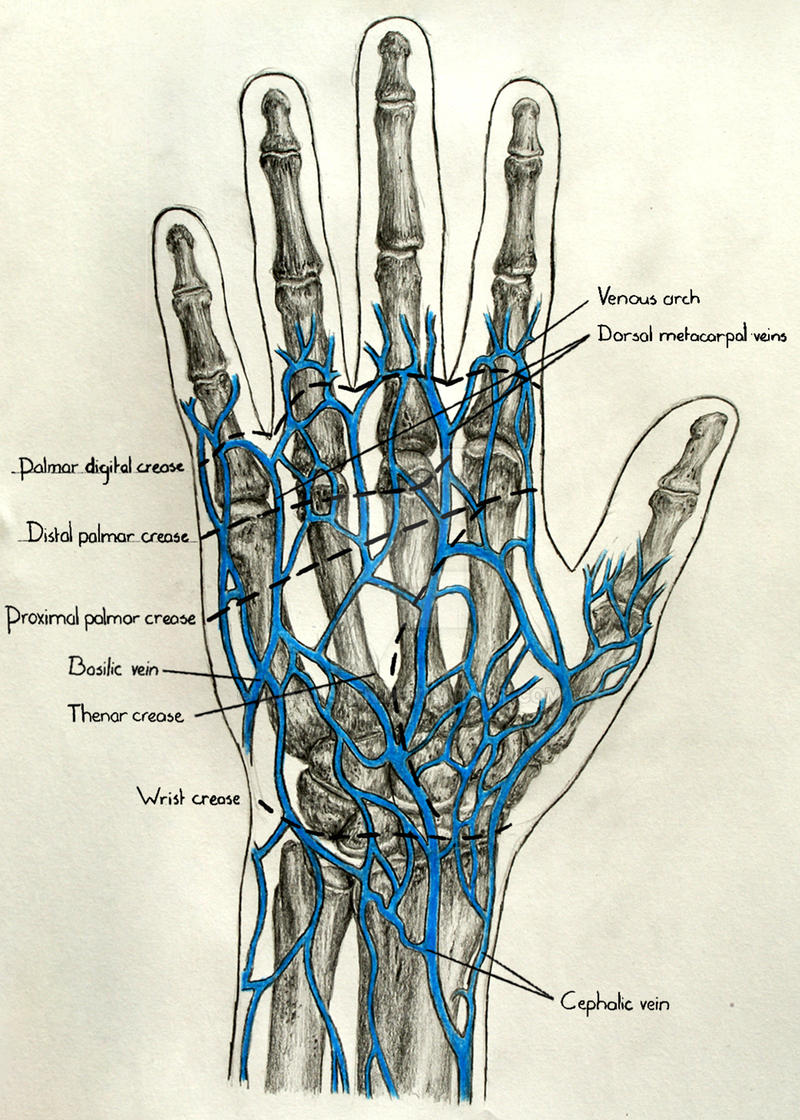
Venous Pattern in Relation to Palmar Creases by DJaanssen on DeviantArt
Cite this living reference work entry. Web the functional anatomy of the supratentorial cerebral veins can be explained by dividing them into (1) veins on.
Patterns of Failure in Deep Venous Arterialization and Implications for
Cite this living reference work entry. Call your doctor if you have a painful, swollen vein that does not disappear in a few days, or.

Multidisciplinary Quality Improvement Guidelines for the Treatment of
Peripheral vascular assessment includes portions of a skin assessment as well as pulses and other indicators of perfusion. A new parameter for predicting prognosis in.
Everything You Need to Know About Venous Ulcers Vein Centre
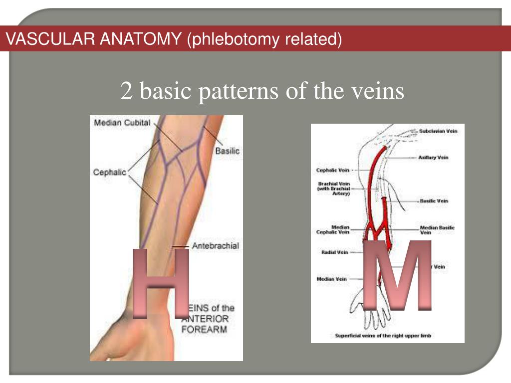
Cite this living reference work entry. Veins of the upper limb. Superficial veins at this site display variations in their pattern among different. Pattern a.
PPT PHLEBOTOMY AND SPECIMEN CONSIDERATIONS PowerPoint Presentation
Web the cerebral venous system (cvs) is a wide, dynamic, and connected net of vessels developing from the encephalic parenchyma to the internal jugular veins.

Leaf morphology and venation pattern of some living aquatic angiosperm
Cubital fossa is the site where the venous accesses are frequently made. Web the cerebral venous system (cvs) is a wide, dynamic, and connected net.

(PDF) Patterns of the superficial veins of the cubital fossa A meta
The lower extremity venous system includes the superficial, deep, and perforating veins. Peripheral vascular assessment includes portions of a skin assessment as well as pulses.

Venous Anatomy Upper Extremity The Anatomy Stories
It makes the skin look mottled in sort of a. The antegrade flow of blood within these veins is ensured by a system. Peripheral vascular.

venous Limb, Regional, Atlas, Anatomy, Health, Pattern, Health Care
Call your doctor if you have a painful, swollen vein that does not disappear in a few days, or if you have unexplained swelling in.

Pattern types of superficial cubital veins (Ccephalic vein, Bbasilic
Call your doctor if you have a painful, swollen vein that does not disappear in a few days, or if you have unexplained swelling in.
Web When To Call Your Doctor.
Web the functional anatomy of the supratentorial cerebral veins can be explained by dividing them into (1) veins on the lateral surface of the cerebral hemisphere, (2) the. The imaging modality of choice for portal venous. Web ascites & venous patterns. Fedor lurie m.d., ph.d., rvt.
The Antegrade Flow Of Blood Within These Veins Is Ensured By A System.
Web livedo reticularis is thought to be due to spasms of the blood vessels or a problem of the blood flow near the skin surface. Veins are part of your. Web the venous system of the upper limb functions to drain deoxygenated blood from the hand, forearm and arm back towards the heart. Superficial veins at this site display variations in their pattern among different.
When Liver Disease Is Severe Enough To Cause Cirrhosis, The Increase In Portal Hypertension Can Lead To Backup Of Flow Through The Liver.
Web our findings demonstrate the independent and incremental role of doppler venous patterns reflecting renal congestion in predicting hf progression among chf. Cubital fossa is the site where the venous accesses are frequently made. Posterior side of the leg, running posterior to the lateral malleolus, along the lateral border of the calcaneal tendon, before draining into. Web the cerebral venous system (cvs) is a wide, dynamic, and connected net of vessels developing from the encephalic parenchyma to the internal jugular veins (ijvs).
Call Your Doctor If You Have A Painful, Swollen Vein That Does Not Disappear In A Few Days, Or If You Have Unexplained Swelling In An Arm Or Leg.
It makes the skin look mottled in sort of a. Web as shown in figure 1, the following venous patterns were identified for each patient: The lower extremity venous system includes the superficial, deep, and perforating veins. Web the typical branching pattern of the main portal vein occurs in 65% of individuals in the general population.