Strain Pattern Ecg - This ecg* demonstrates a strain pattern isolated to v5 and v6. Typically, these ecgs come from ats category 2 patients that have been triaged but not yet seen by a. Web lvh with strain pattern can sometimes be seen in long standing severe aortic regurgitation, usually with associated left ventricular hypertrophy and systolic dysfunction. Web left ventricular hypertrophy (lvh): The inner lining (endocardium), a thick muscle layer (myocardium) and an outer lining (epicardium).the myocardium is the thick muscle layer, in which the muscle fibers are organized into several sheets. Number one cause of sudden cardiac death in. The two most common pressure overload states are systemic hypertension and aortic stenosis. The patient had severe concentric lvh by echo, but no ecg voltage criteria for lvh. 2,6 ecg strain has been. Web left ventricular hypertrophy (lvh) refers to an increase in the size of myocardial fibers in the main cardiac pumping chamber.
.jpg)
ECG Interpretation ECG Interpretation Review 51 (Chamber Enlargement
Web lvh with strain pattern can sometimes be seen in long standing severe aortic regurgitation, usually with associated left ventricular hypertrophy and systolic dysfunction. Inferior.

STT wave appearance of normal (A) — vs “strain” (C) GrepMed
This pattern is associated with high pulmonary artery pressures (34%) right axis deviation (16%). However, the independence of the relation of strain to increased lv.

The ECG in left ventricular hypertrophy (LVH) criteria and
Web 3:44 pm pdt. Asymptomatic patients with nf1 had normal electrocardiograms, none with the typical ecg patterns described in ns. [1] it is an abnormality.
Right Heart Strain ECG Stampede
Number one cause of sudden cardiac death in. The strain pattern in the 12‐lead ecg, defined as st‐segment depression and t‐wave inversion, represents ventricular repolarization.

Left Ventricular Hypertrophy LVH with Strain Pattern on ECG YouTube
Web 3:44 pm pdt. Web left ventricular hypertrophy (lvh): 1 the mechanism underlying ecg strain is unclear, although it has been proposed as subendocardial ischemia..
Strain pattern ecardiogram
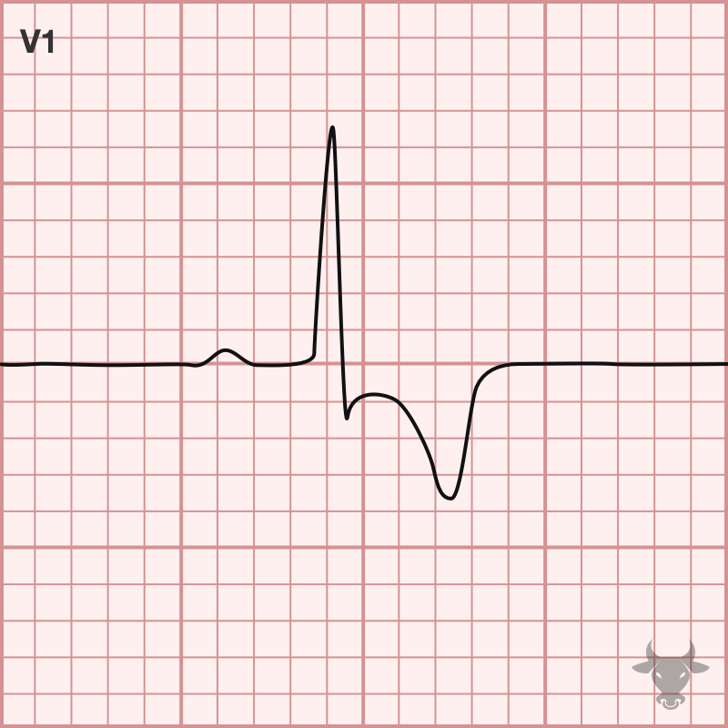
Prevalence ~1 in 500 people. R/s ratio in v1 > 1. Web 3:44 pm pdt. However, the independence of the relation of strain to increased.
Right Ventricular Strain • LITFL • ECG Library Diagnosis
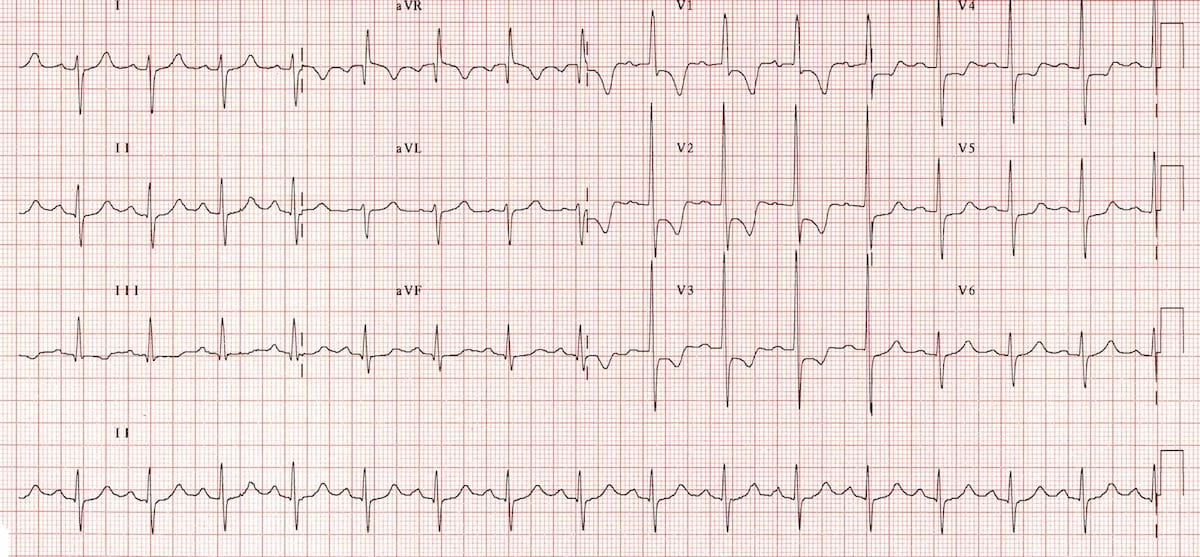
7 indeed, the presence of even minimal degrees of. In addition, classic voltage criteria for lvh are present—cornell criteria >28 mm in ravl (19 mm).
Strain, strain rate and speckle tracking Myocardial deformation ECG
Peer reviewed with thanks by dr stephen smith of dr smith’s ecg blog. Web left ventricular hypertrophy (lvh): However, the independence of the relation of.

ECG Essentials Ischemia & Ventricular Strain YouTube
However, the independence of the relation of strain to increased lv mass from its relation to chd has not been extensively examined. Asymptomatic patients with.
PPT ECG PRACTICAL APPROACH PowerPoint Presentation, free download
In addition, classic voltage criteria for lvh are present—cornell criteria >28 mm in ravl (19 mm) and s v3 12 mm—along. Huge precordial r and.
Huge Precordial R And S Waves That Overlap With The Adjacent Leads (Sv2 + Rv6 >> 35 Mm).
Moreover, ecg strain was associated with increased myocardial injury and impaired left ventricular performance and was an independent. [1] it is an abnormality of repolarization and it has been associated with an adverse prognosis in a variety heart disease patients. Web right ventricular strain is a repolarisation abnormality due to right ventricular hypertrophy (rvh) or dilatation. This ecg* demonstrates a strain pattern isolated to v5 and v6.
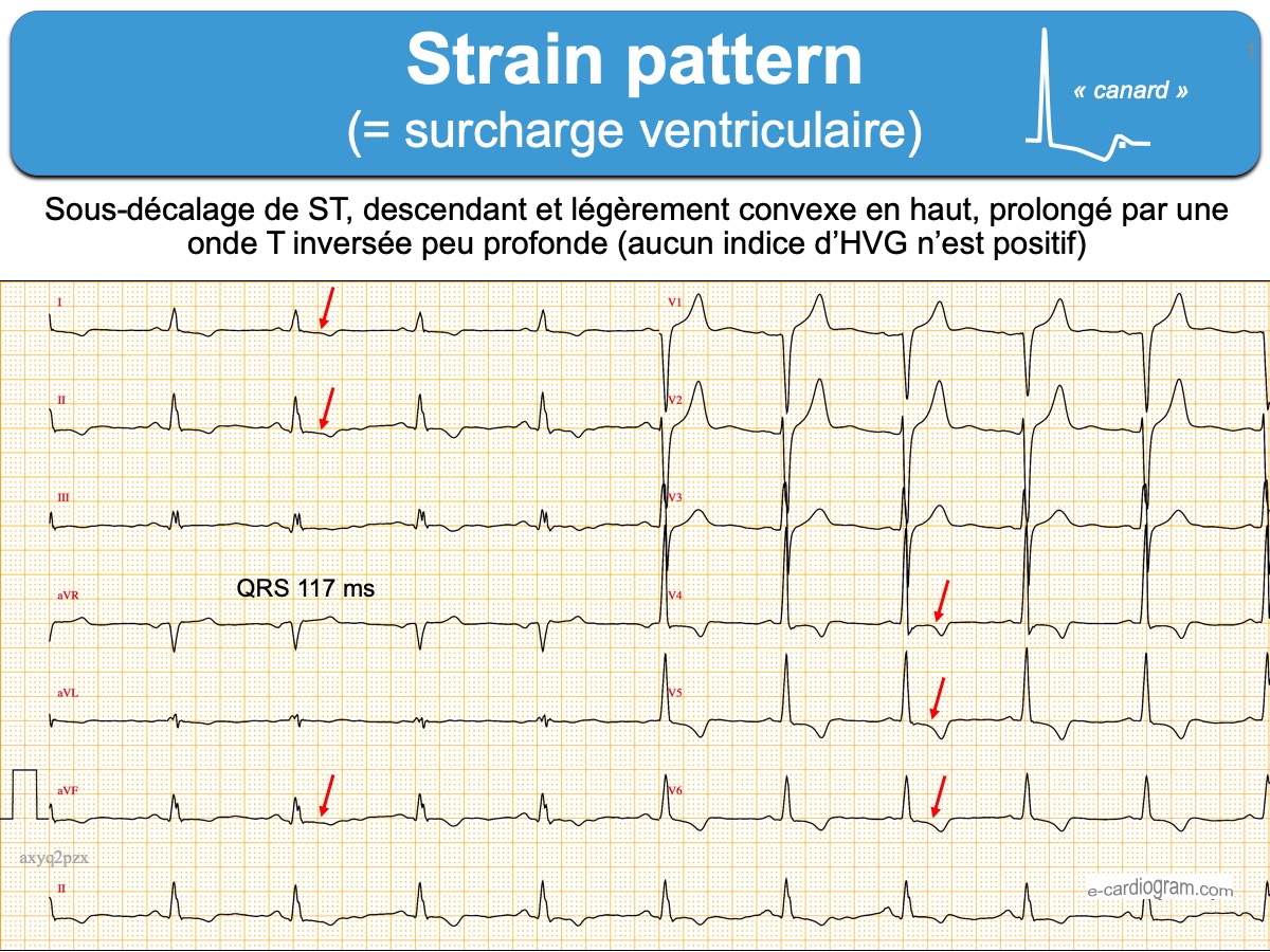
The Strain Pattern In The 12‐Lead Ecg, Defined As St‐Segment Depression And T‐Wave Inversion, Represents Ventricular Repolarization Abnormalities.1 The Mechanism Underlying Ecg Strain Is Unclear, Although It Has Been Proposed As Subendocardial Ischemia.2, 3 Ecg Strain Is Associated With Concentric Left Ventricular.
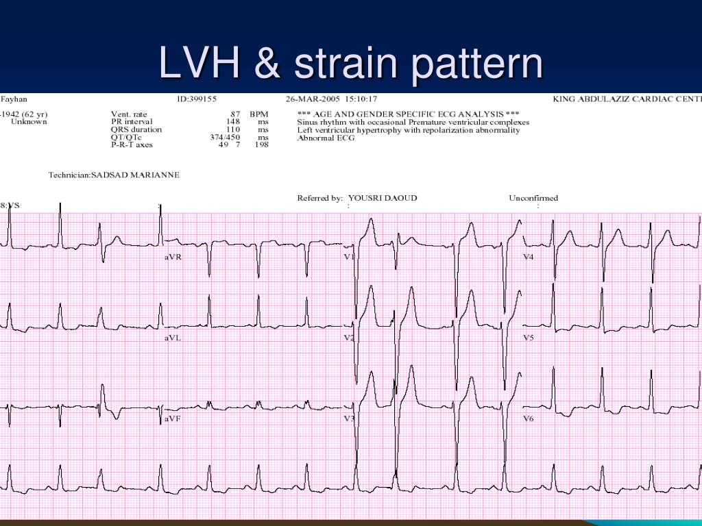
Mets manager carlos mendoza said sunday. St depression and t wave inversion in leads corresponding to the right ventricle: Hypertrophic cardiomyopathy, previously termed hypertrophic obstructive cardiomyopathy (hocm) or idiopathic hypertrophic subaortic stenosis (ihss), is one of the most common inherited cardiac disorders: The sensitivity of lvh strain pattern on ecg as a measure of lvh has ranged from 3.8% to 50% in various reports [1].
1 The Mechanism Underlying Ecg Strain Is Unclear, Although It Has Been Proposed As Subendocardial Ischemia.
Web this ecg* demonstrates strain pattern in leads i, avl, v5, and v6. Typically, these ecgs come from ats category 2 patients that have been triaged but not yet seen by a. R/s ratio in v1 > 1. This pattern is associated with high pulmonary artery pressures (34%) right axis deviation (16%).
Inferior Leads Ii, Iii, Avf, Often Most Pronounced In Lead Iii As This Is The Most Rightward Facing.
Web left ventricular hypertrophy (lvh): Strain, strain rate, speckle tracking. Web these ecg changes were previously referred to as strain pattern because it was believed that they indicated left ventricular exhaustion. The patient had severe concentric lvh by echo, but no ecg voltage criteria for lvh.