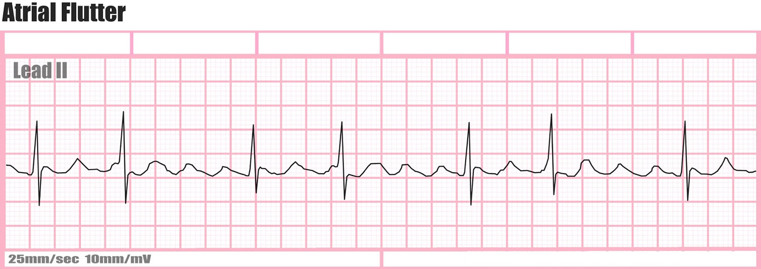
Saw Tooth Pattern Ecg - After questioning him, it was clear that he had been having palpitations and near dizzy spells on and off for a couple of weeks. Web the ecg pattern may not reflect the mechanism. By observing the inferior leads we can determine the direction of the stimulus and classify it further as counterclockwise atrial flutter or clockwise atrial flutter. Web on the surface electrocardiogram, the atrial flutter wave forms a “sawtooth” pattern in the inferior leads ( fig. 1 the ecg in type i (typical) afl is characterized by an inverted sawtooth flutter (f) wave pattern in. Web ecg strip (electrocardiogram, ekg) of sawtooth pattern of atrial flutter. If atrial flutter is associated with a 2:1 block, p waves are difficult to recognise because of the superposition of the preceding t waves. The initial descent is gradual, followed by a sharp, steep component. (a) ecg fulfilling classical flutter criteria (rate and lack of isoelectric baseline) in a case of focal tachycardia originating in the right superior pulmonary vein. The foci responsible for focal atrial tachycardia do not occur randomly throughout the atria but tend to cluster at characteristic anatomical locations.
PPT ECG Basics Module 2( Arrhythmias level 1) PowerPoint Presentation
Web on the surface electrocardiogram, the atrial flutter wave forms a “sawtooth” pattern in the inferior leads ( fig. Atrial flutter is the only diagnosis.
![Atrial Flutter ECG Interpretation [With Examples] Manual of Medicine](https://manualofmedicine.com/wp-content/uploads/2022/01/Atrial-Flutter-with-Variable-AV-Block.png)
Atrial Flutter ECG Interpretation [With Examples] Manual of Medicine
By observing the inferior leads we can determine the direction of the stimulus and classify it further as counterclockwise atrial flutter or clockwise atrial flutter..
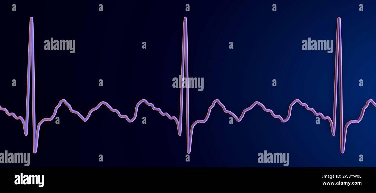
Illustration depicting an atrial flutter abnormal heart rhythm on an
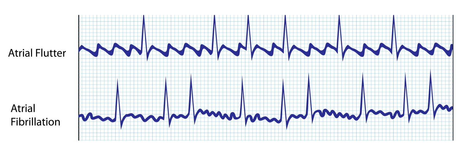
Web atrial flutter produces a distinctive “sawtooth” pattern on an electrocardiogram (ekg or ecg), a test used to monitor the heart and diagnose heart rhythm.
Atrial Flutter Symptoms, Causes, and Treatment Dr. AFib
Three findings that distinguish atypical atrial flutter arrhythmia from typical atrial flutter: Web the electrocardiogram shows a saw tooth's pattern in inferior leads, with a.

Electrocardiogram showing the ''sawtooth'' or ''picket fence'' pattern
I admitted him to the hospital and started a blood thinner and an intravenous drug to slow his heart rate. Note the irregular ventricular rate..
Atrial Flutter Causes, Symptoms, Treatment & Ablation
If atrial flutter is associated with a 2:1 block, p waves are difficult to recognise because of the superposition of the preceding t waves. The.
![Atrial Flutter ECG Interpretation [With Examples] Manual of Medicine](https://manualofmedicine.com/wp-content/uploads/2022/01/Atrial-Flutter-with-2-1-AV-Block.png)
Atrial Flutter ECG Interpretation [With Examples] Manual of Medicine
Web the electrocardiographic pattern of typical ccwid flutter waves is easily recognizable: Upright flutter waves in v1 that may resemble p waves; Three findings that.

Arrhythmias Originating in the Atria Cardiac Electrophysiology Clinics
Web on the surface electrocardiogram, the atrial flutter wave forms a “sawtooth” pattern in the inferior leads ( fig. The initial descent is gradual, followed.

Electrocardiogram showing the ''sawtooth'' or ''picket fence'' pattern
I admitted him to the hospital and started a blood thinner and an intravenous drug to slow his heart rate. 1 the ecg in type.

Electrocardiogram showing typical atrial flutter with variable
1 the ecg in type i (typical) afl is characterized by an inverted sawtooth flutter (f) wave pattern in. By observing the inferior leads we.
(A) Ecg Fulfilling Classical Flutter Criteria (Rate And Lack Of Isoelectric Baseline) In A Case Of Focal Tachycardia Originating In The Right Superior Pulmonary Vein.
Web typical atrial flutter has been described as counterclockwise reentry within right atrial and it presents a characteristic ecg “sawtooth” pattern on the inferior leads. Web atrial flutter produces a distinctive “sawtooth” pattern on an electrocardiogram (ekg or ecg), a test used to monitor the heart and diagnose heart rhythm disorders. After questioning him, it was clear that he had been having palpitations and near dizzy spells on and off for a couple of weeks. Conducted with right bundle branch block.) carotid sinus massage can increase av block and better expose the typical flutter waves.
Last Reviewed 1 Jan 2018.
Web the electrocardiographic pattern of typical ccwid flutter waves is easily recognizable: Catheter ablation is highly successful and is considered the definitive treatment for typical atrial. Upright flutter waves in v1 that may resemble p waves; Web atrial flutter (afl) is a cardiac dysrhythmia characterized by rapid and regular depolarization of the atria that appears as a sawtooth pattern on the electrocardiogram (ecg) and is categorized into type i (typical) and type ii (atypical) afl.
Intracardiac Mapping Has Shown What.
How to differentiate atypical flutter from typical flutter? Web sawtooth pattern in ecg atrial flutter (afl) may exist with or without underlying structural heart disease. The electrical signal that causes atrial flutter (afl) circulates in an organized, predictable pattern. Web in typical flutter, ecg shows continuous and regular atrial activation with a sawtooth pattern, most obvious in leads ii, iii, and avf (see figure atrial flutter ).
Web On An Ecg, Atrial Flutter Resembles F Waves With A Sawtooth Pattern.
The initial descent is gradual, followed by a sharp, steep component. Web the ecg pattern may not reflect the mechanism. If atrial flutter is associated with a 2:1 block, p waves are difficult to recognise because of the superposition of the preceding t waves. Electrocardiographic approach to atrial flutter.