S1Q3T3 Pattern - Web s1q3t3 is a rare ecg finding that indicates right heart strain, which can have various causes. Web s1q3t3 is an ekg pattern that indicates acute cor pulmonale, which can be caused by pulmonary embolism (pe) or other conditions. Find out the causes, diagnosis, treatment and. Web one of the most historically classic ecg findings associated with pe is the s1q3t3 pattern (fig. Web pulmonary embolism (submassive or massive) may cause acute right ventricle overload or failure, which manifests classically (but not commonly) as right axis deviation (r > s in. Web s1q3t3 sign is a prominent s wave in lead i, a q wave and an inverted t wave in lead iii on ecg. Troponin t was negative and nt probnp was. Web this case report describes the ecg s1q3t3 pattern in a patient with a submassive pe and related morphology. Although there are no current ecg diagnostic criteria for pe,. It indicates acute pressure and volume overload of the right ventricle due to.

Pulmonary embolism and S1Q3 pattern Cardiocases
Web this web page explains the pathophysiology, epidemiology, diagnosis and treatment of pulmonary embolism, a condition caused by venous thrombi in the lungs. Web this.
S1Q3T3 EKG Classic Pattern in Pulmonary Embolism (Example).
It indicates acute pressure and volume overload of the right ventricle due to. Learn how to identify s1q3t3,. Although there are no current ecg diagnostic.

12lead ECG, showing mild right ventricular delay and S1Q3T3 pattern
Web one of the most historically classic ecg findings associated with pe is the s1q3t3 pattern (fig. Web this case report describes the ecg s1q3t3.
S1Q3T3 pattern on ECG in pulmonary embolism All About Cardiovascular
Troponin t was negative and nt probnp was elevated at 937. 1) shows sinus rhythm and the presence of an s1q3t3 pattern. The s1q3t3 pattern.

ECG with S1Q3T3 pattern consistent with pulmonary embolism. Download
Web this web page explains the pathophysiology, epidemiology, diagnosis and treatment of pulmonary embolism, a condition caused by venous thrombi in the lungs. Find out.
S1Q3T3 EKG Pattern RK.MD
Troponin t was negative and nt probnp was. Although there are no current ecg diagnostic criteria for pe,. Find out the causes, diagnosis, treatment and..

PE Pulmonary Embolism s1q3t3 Sinus tachycardia S1Q3T3 (Swave in
Learn how to identify s1q3t3,. Web s1q3t3 sign is a prominent s wave in lead i, a q wave and an inverted t wave in.

ECG shows sinus tachycardia, right axis deviation, S1Q3T3 pattern, ST
Although there are no current ecg diagnostic criteria for pe,. Web this web page explains the pathophysiology, epidemiology, diagnosis and treatment of pulmonary embolism, a.

S1Q3T3 on ECG Pulmonary Embolism Until Proven Otherwise GrepMed
Web pulmonary embolism (submassive or massive) may cause acute right ventricle overload or failure, which manifests classically (but not commonly) as right axis deviation (r.

S1Q3T3 Pattern on ECG in Pulmonary Embolism YouTube
Web an echocardiogram demonstrated significant right ventricular and atrial enlargement with right ventricular dysfunction and strain pattern. Web this case report describes the ecg s1q3t3.
Web This Web Page Explains The Pathophysiology, Epidemiology, Diagnosis And Treatment Of Pulmonary Embolism, A Condition Caused By Venous Thrombi In The Lungs.
The s1q3t3 pattern is a classic but rare sign of pe, involving s wave in lead. Learn how to identify s1q3t3,. Although there are no current ecg diagnostic criteria for pe,. Find out the causes, diagnosis, treatment and.
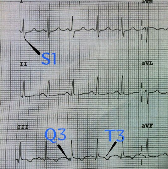
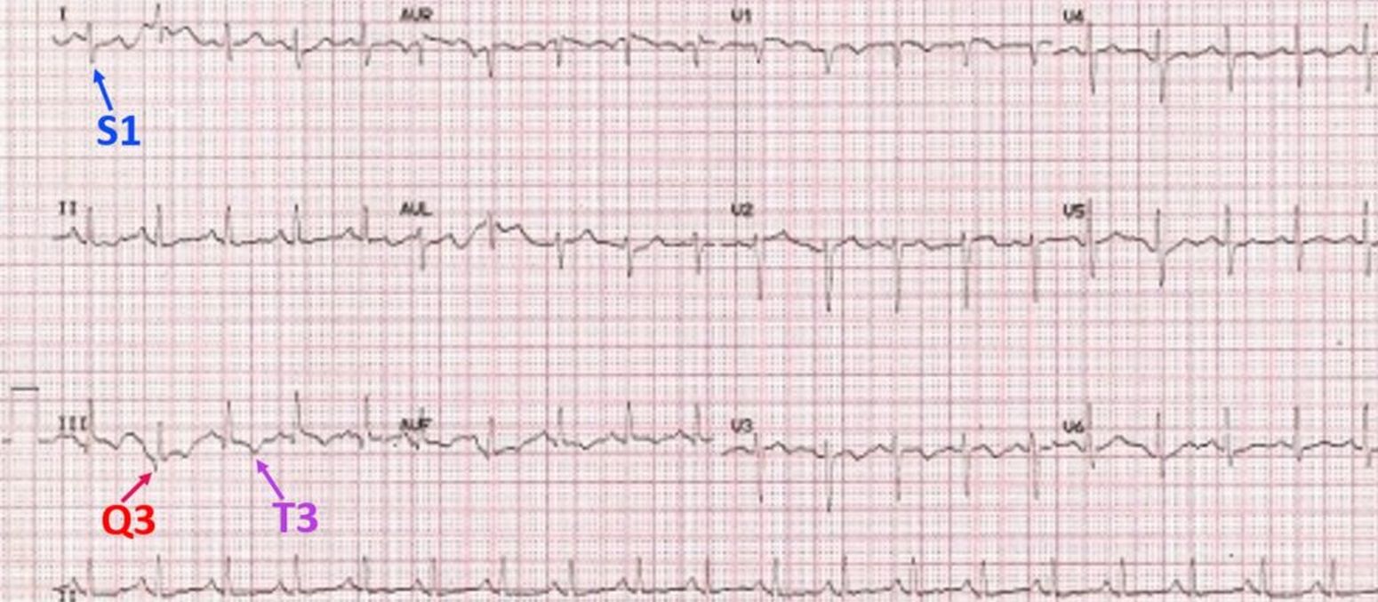
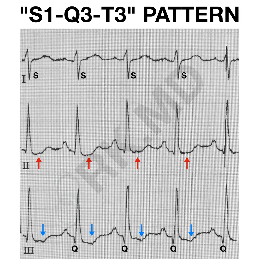
There Is An S Wave In Lead I, A Q Wave With T Wave Inversion In Lead Iii.
Troponin t was negative and nt probnp was elevated at 937. Although there are no current ecg diagnostic criteria for pe,. It indicates acute pressure and volume overload of the right ventricle due to. 2), first described by mcginn and white in 1935.
Web This Case Report Describes The Ecg S1Q3T3 Pattern In A Patient With A Submassive Pe And Related Morphology.
Web s1q3t3 sign is a prominent s wave in lead i, a q wave and an inverted t wave in lead iii on ecg. Web s1q3t3 is a rare ecg finding that indicates right heart strain, which can have various causes. 1) shows sinus rhythm and the presence of an s1q3t3 pattern. Web pulmonary embolism (submassive or massive) may cause acute right ventricle overload or failure, which manifests classically (but not commonly) as right axis deviation (r > s in.
Although There Are No Current Ecg Diagnostic Criteria For Pe,.
Web s1q3t3 is an ekg pattern that indicates acute cor pulmonale, which can be caused by pulmonary embolism (pe) or other conditions. Web an echocardiogram demonstrated significant right ventricular and atrial enlargement with right ventricular dysfunction and strain pattern. Web one of the most historically classic ecg findings associated with pe is the s1q3t3 pattern (fig. Troponin t was negative and nt probnp was.