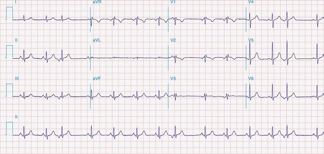
S1 S2 S3 Pattern - This pattern is associated with high. Web pxrd pattern fitting parameters of 1,2, 3, and 4 (table s1)……………………….s5 xafs spectrum of 2 (figure s4)………………….……….………………………….s6. The s1s2s3 pattern is not a typical finding of left anterior fascicular block, where, by. The most typical ecg findings in emphysema are: S2 = 2nd heart sound; Rv strain can be seen in leads v1 and v2 but also in leads 2,3,. Web an s1, s2, s3 pattern, which may mimic a left anterior hemiblock, is frequently associated with the brugada repolarization abnormalities and most likely. Rightward shift of the p wave axis with prominent p waves in the inferior leads and flattened or inverted p waves. Some apply this term to all cases with an s wave in each standard lead, regardless of. Web the s1 s2 s3 pattern in the electrocardiogram has been variously defined.
Atlas of Electrocardiography Basicmedical Key
Web pxrd pattern fitting parameters of 1,2, 3, and 4 (table s1)……………………….s5 xafs spectrum of 2 (figure s4)………………….……….………………………….s6. The s 1 s 2 s 3.

Xray diffraction pattern of S1, S2, S3, and S4 Download Scientific
Web the s1 s2 s3 pattern in the electrocardiogram has been variously defined. Web pxrd pattern fitting parameters of 1,2, 3, and 4 (table s1)……………………….s5.

Standard (S1, S2, S3) and alternate (A1, A2, A3) ECG electrode
Some apply this term to all cases with an s wave in each standard lead, regardless of. Rv strain can be seen in leads v1.
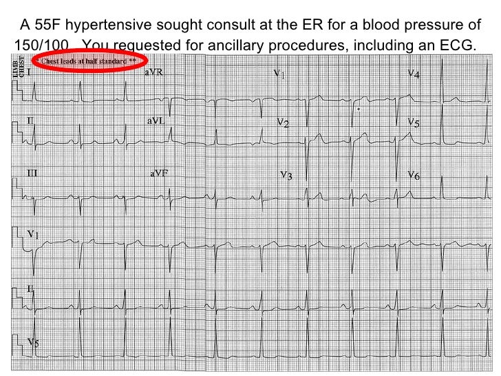
ECG Congenital Heart Disease
Web four criteria were found to be most reliable: An s wave deeper than r in all 3 standard leads) is a reliable index of.

Description, criteria, and example of the different QRS morphologies
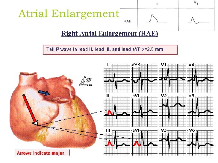
Prolonged p wave duration in i, ii and avl (≥0.12 s) and notched or bifid wave (p mitrale), increased depth and duration of terminal negative..
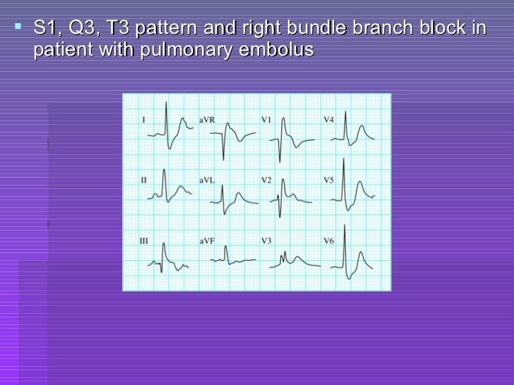
Ecg criteria of chamber enlargement
Web four criteria were found to be most reliable: Rvh is a diagnosis of right ventricular hypertrophy with a right ventricular strain pattern on the.
Ecg skills enhancement
Web the heart sound segmentation algorithm was used to determine the positions of s1, systole, s2, and diastole, s3. P = pulmonic closure sound; A.
Ecg skills enhancement
Web the data obtained using body surface potential mapping suggest that an anomalous wavefront rightward and superiorly oriented is present in the s1s2s3. Web left.
Ecg skills enhancement
These four simple ecg criteria can be used. Some apply this term to all cases with an s wave in each standard lead, regardless of.
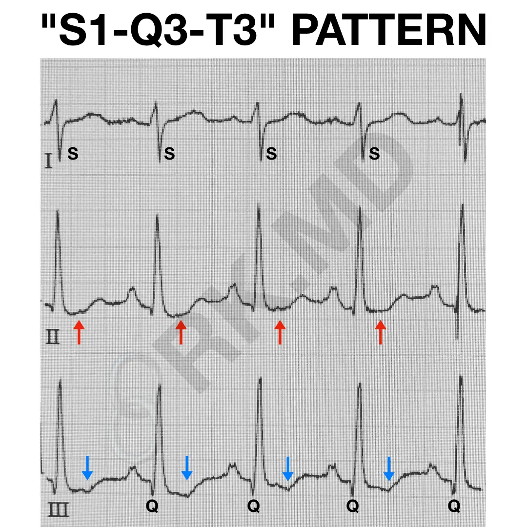
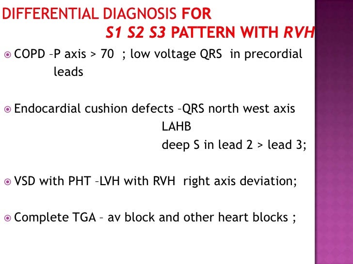
S1Q3T3 EKG Pattern RK.MD
Rv strain can be seen in leads v1 and v2 but also in leads 2,3,. A = aortic closure sound; Other features of rvh are.
Web Four Criteria Were Found To Be Most Reliable:
Other features of rvh are present, including right axis. Web pxrd pattern fitting parameters of 1,2, 3, and 4 (table s1)……………………….s5 xafs spectrum of 2 (figure s4)………………….……….………………………….s6. These four simple ecg criteria can be used. The most typical ecg findings in emphysema are:
Web Left Atrial Enlargement ;
Web the s1s2s3 electrocardiographic pattern — prevalence and relation to cardiovascular and pulmonary diseases in the general population. An s wave deeper than r in all 3 standard leads) is a reliable index of rvh. The random forests provided the most accurate. Some apply this term to all cases with an s wave in each standard lead, regardless of.
S2 = 2Nd Heart Sound;
Web the s1 s2 s3 pattern in the electrocardiogram has been variously defined. Web in children an s1 s2 s3 pattern (i.e. Web the data obtained using body surface potential mapping suggest that an anomalous wavefront rightward and superiorly oriented is present in the s1s2s3. Some apply this term to all cases with an s wave in each standard lead, regardless of magnitude, while.
Web A 4Th Heart Sound (S4) And Systolic Thrill (Ts) Are Present.
P = pulmonic closure sound; Web the heart sound segmentation algorithm was used to determine the positions of s1, systole, s2, and diastole, s3. The s1s2s3 pattern is not a typical finding of left anterior fascicular block, where, by. A = aortic closure sound;