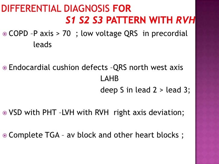
S1 S2 S3 Pattern On Electrocardiogram - Prolonged p wave duration in i, ii and avl (≥0.12 s) and notched or bifid wave (p mitrale), increased depth and duration of terminal. Web an s1, s2, s3 pattern, which may mimic a left anterior hemiblock, is frequently associated with the brugada repolarization abnormalities and most likely. Web the s 1 s 2 s 3 pattern in the electrocardiogram has been variously defined. Web a 4th heart sound (s4) and systolic thrill (ts) are present. Web typical ecg findings in copd. Web left atrial enlargement ; Web the s1s2s3 electrocardiographic pattern — prevalence and relation to cardiovascular and pulmonary diseases in the general population. Web the data obtained using body surface potential mapping suggest that an anomalous wavefront rightward and superiorly oriented is present in the s1s2s3 pattern, which is. Web biatrial enlargement is diagnosed when criteria for both right and left atrial enlargement are present on the same ecg. Web in the general adult population, the prevalence of the s1s2s3 ecg pattern is markedly affected by the diagnostic ecg criteria.

Description, criteria, and example of the different QRS morphologies
Web the s1s2s3 sign has been associated with pulmonary embolism, chronic obstructive pulmonary disease (copd), and obstructive sleep apnea and with right ventricular. Web left.
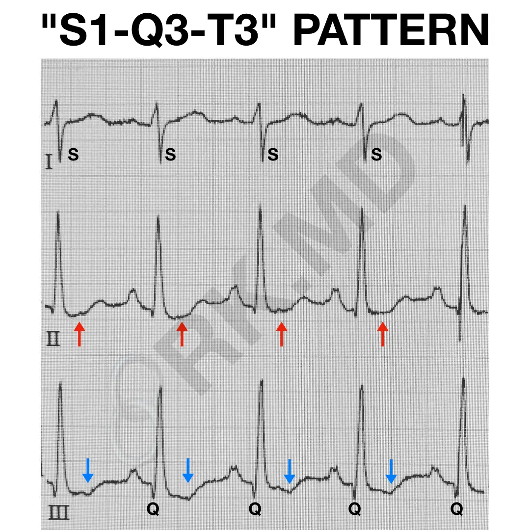
S1Q3T3 EKG Pattern RK.MD
S2 = 2nd heart sound; The most typical ecg findings in emphysema are: A = aortic closure sound; Web the s1 s2 s3 pattern in.

Heart Sounds Diagram S1 S2
Prolonged p wave duration in i, ii and avl (≥0.12 s) and notched or bifid wave (p mitrale), increased depth and duration of terminal. Web.

Standard (S1, S2, S3) and alternate (A1, A2, A3) ECG electrode
A = aortic closure sound; The s 1 s 2 s 3 pattern in the electrocardiogram has been variously defined. Web the s1s2s3 sign has.
ECG Congenital Heart Disease
P = pulmonic closure sound; Some apply this term to all cases with an s wave in each standard lead, regardless of magnitude,. Hcm (septal.
Figure 15. Cardiac Rhythm Interpretation
The diagnosis of biatrial enlargement. Some apply this term to all cases with an s wave in each standard lead, regardless of magnitude,. Web an.

Heart Sounds Diagram S1 S2
Web s1 s2 s3 pattern in children. Web a 4th heart sound (s4) and systolic thrill (ts) are present. S1 = 1st heart sound; Web.
Atlas of Electrocardiography Basicmedical Key
Web an s1, s2, s3 pattern, which may mimic a left anterior hemiblock, is frequently associated with the brugada repolarization abnormalities and most likely. Web.

【コラム051】S1S2S3パターンを考えます。 Cardio2012のECGブログ2019改
Some apply this term to all cases with an s wave in each standard lead, regardless of. Some apply this term to all cases with.

PE Pulmonary Embolism s1q3t3 Sinus tachycardia S1Q3T3 (Swave in
Web the data obtained using body surface potential mapping suggest that an anomalous wavefront rightward and superiorly oriented is present in the s1s2s3 pattern, which.
Some Apply This Term To All Cases With An S Wave In Each Standard Lead, Regardless Of Magnitude,.
S1 = 1st heart sound; Web s1 s2 s3 pattern in children. Web typical ecg findings in copd. The s 1 s 2 s 3 pattern in the electrocardiogram has been variously defined.
Web In The General Adult Population, The Prevalence Of The S1S2S3 Ecg Pattern Is Markedly Affected By The Diagnostic Ecg Criteria.
P = pulmonic closure sound; Web left atrial enlargement ; Some apply this term to all cases with an s wave in each standard lead, regardless of. A = aortic closure sound;
Rightward Shift Of The P Wave Axis With Prominent P Waves In The.
Prolonged p wave duration in i, ii and avl (≥0.12 s) and notched or bifid wave (p mitrale), increased depth and duration of terminal. Web an s1, s2, s3 pattern, which may mimic a left anterior hemiblock, is frequently associated with the brugada repolarization abnormalities and most likely. Web the s1s2s3 sign has been associated with pulmonary embolism, chronic obstructive pulmonary disease (copd), and obstructive sleep apnea and with right ventricular. Web the s1 s2 s3 pattern in the electrocardiogram has been variously defined.
Web A 4Th Heart Sound (S4) And Systolic Thrill (Ts) Are Present.
Web the data obtained using body surface potential mapping suggest that an anomalous wavefront rightward and superiorly oriented is present in the s1s2s3. The diagnosis of biatrial enlargement. Hcm (septal hypertrophy) kulbertus' block (septal fascicular block) duchennes muscular. Web the data obtained using body surface potential mapping suggest that an anomalous wavefront rightward and superiorly oriented is present in the s1s2s3 pattern, which is.