S1 S2 S3 Pattern In Ecg - Note that there is a prominent s wave in leads 1, 2, and 3 and the s waves are equal in duration and magnitude to the preceding r waves. Web the s1 s2 s3 pattern in the electrocardiogram has been variously defined. Some apply this term to all cases with an s wave in each standard lead, regardless of magnitude, while others use it to indicate situations where the prominent qrs deflection is. Other abnormalities caused by rvh An amplitude of at least 1.5 mm — was found in 423 subjects (6.7%). Web the s1s2s3 pattern has had variable criteria for identifying rv dysfunction and pulmonary disease since its initial description in 1960 by burch and de pasquale in association with ventricular septal defect. Web right ventricular strain is a repolarisation abnormality due to right ventricular hypertrophy (rvh) or dilatation. Rv strain can be seen in leads v1 and v2 but also in leads 2,3, avf. An s wave deeper than r in all 3 standard leads) is a reliable index of rvh. Ekg in right ventricular hypertrophy.

Heart Sounds Diagram S1 S2
Deep s waves in the left precordial leads v 5 and v 6 (r:s <1); Inferior leads ii, iii, avf, often most pronounced in lead.

【コラム051】S1S2S3パターンを考えます。 Cardio2012のECGブログ2019改
Of these, 80 subjects (18.9%) had an associated s2 ≥ s3 pattern. Web ecg changes occur in chronic obstructive pulmonary disease (copd) due to: Ecg.

Description, criteria, and example of the different QRS morphologies
Web right ventricular strain is a repolarisation abnormality due to right ventricular hypertrophy (rvh) or dilatation. Some apply this term to all cases with an.

The Electrocardiogram explained What is an ECG?
In some cases s waves are equal or superior to r waves in one or more limb leads.' Web the s1 s2 s3 pattern in.
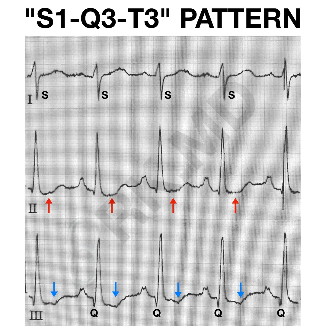
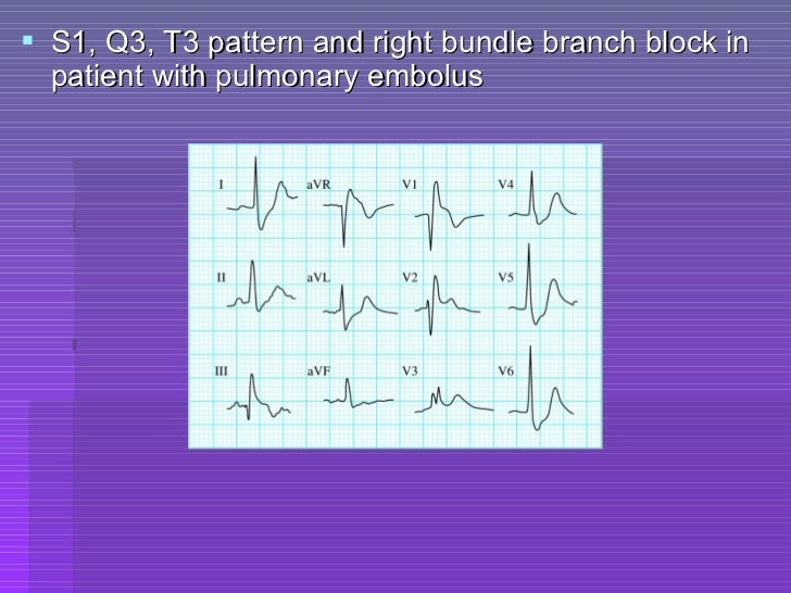
S1Q3T3 EKG Pattern RK.MD
Web an s1, s2, s3 pattern, which may mimic a left anterior hemiblock, is frequently associated with the brugada repolarization abnormalities and most likely reflects.
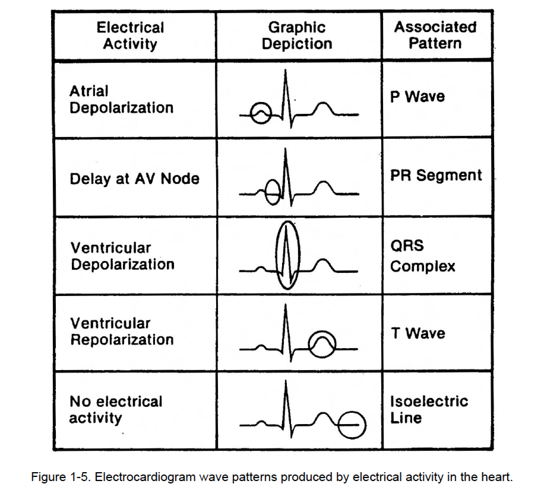
Figure 15. Cardiac Rhythm Interpretation
Definition of diseases classification of coronary heart disease (chd) required at least one 6, june, 1974 ecg diagnosis of right ventricular hypertrophy in. Web in.
Atlas of Electrocardiography Basicmedical Key
Some apply this term to all cases with an s wave in each standard lead, regardless of magnitude, while others use it to indicate situations.

Standard (S1, S2, S3) and alternate (A1, A2, A3) ECG electrode
Web the s1 s2 s3 pattern in the electrocardiogram has been variously defined. Web the chosen criteria have major impact on the prevalence of the.
Ecg criteria of chamber enlargement
His electrocardiogram is an excellent example of the s1, s2, s3 syndrome. Web the purpose of this chapter is to review the role of the.
ECG Congenital Heart Disease
Other abnormalities caused by rvh Some apply this term to all cases with an s wave in each standard lead, regardless of magnitude, while others.
Deep S Waves In The Left Precordial Leads V 5 And V 6 (R:s <1);
Rv strain can be seen in leads v1 and v2 but also in leads 2,3, avf. 6, june, 1974 ecg diagnosis of right ventricular hypertrophy in. The diagnosis of biatrial enlargement requires criteria for lae and rae to be met in either lead ii, lead v1 or a combination of leads. St depression and t wave inversion in leads corresponding to the right ventricle:
Note That There Is A Prominent S Wave In Leads 1, 2, And 3 And The S Waves Are Equal In Duration And Magnitude To The Preceding R Waves.
Rv strain can be seen in leads v1 and v2 but also in leads 2,3, avf; Web in children an s1 s2 s3 pattern (i.e. His electrocardiogram is an excellent example of the s1, s2, s3 syndrome. An amplitude of at least 1.5 mm — was found in 423 subjects (6.7%).
Ecg Criteria For Biatrial Enlargement.
Web biatrial enlargement is diagnosed when criteria for both right and left atrial enlargement are present on the same ecg. Table 4.2 ecg criteria for right. Web the s1s2s3 pattern has had variable criteria for identifying rv dysfunction and pulmonary disease since its initial description in 1960 by burch and de pasquale in association with ventricular septal defect. Some apply this term to all cases with an s wave in each standard lead, regardless of magnitude, while others use it to indicate situations where the prominent qrs deflection is an s wave in these leads.
Web The Purpose Of This Chapter Is To Review The Role Of The Ecg In The Diagnosis Of Cardiac Chamber Enlargement.
In some cases s waves are equal or superior to r waves in one or more limb leads.' Of these, 80 subjects (18.9%) had an associated s2 ≥ s3 pattern. Web with additional ecg criteria; Table 2 shows grouping of various criteria to achieve the maximal sensitivity and specificity range for our patients with copd and rvh.