Rsr Pattern - Web in this review we analyze in detail all the possible conditions, both benign and pathological that may explain the presence of this electrocardiographic pattern. Rbbb pattern in precordial leads. The anatomy, epidemiology, causes, symptoms, ecg findings and diagnosis, differential. Web the brugada syndrome may present with three different ecg patterns, referred to as type 1, type 2, and type 2 brugada syndrome ecg. Web an rsr’ pattern in the right precordial leads is a relatively common electrocardiographic finding that has been described in up to 7% of patients without. Web an rsr’ pattern in the right precordial leads is a relatively common electrocardiographic finding that has been described in up to 7% of patients without. This pattern is associated with high. 3) note appropriate discordance in v1 with st elevation and. An rsr prime (rsr') pattern is a qrs complex with an upward, downward and upward deflection in leads v1 and/or v2. Dominant r wave in v1;
Cureus Transient Giant R Wave as a Marker for Ischemia in Unstable Angina
Web the morphology rsr′ in lead v1: When there is a block in the lbb, the rbb is responsible for spreading the wave. Web in.
Right Bundle Branch Block Exercise Stress Test Online degrees
Web the rsr pattern has also been called “rabbit ears” or an “m” (see right bundle branch block ). Web in other cases, a normal.

Figure 11 from Differential diagnosis of rSr' pattern in leads V1 V2
Web the morphology rsr′ in lead v1: 3) note appropriate discordance in v1 with st elevation and. Comprehensive review and proposed algorithm. Dominant r wave.

(A) ECG showing minimal preexcitation (rsR= pattern in lead III) only
The most typical, and diagnostic, is type. Web the brugada syndrome may present with three different ecg patterns, referred to as type 1, type 2,.

Atrial septal defect electrocardiogram wikidoc
Dominant r wave in v1; The most typical, and diagnostic, is type. Web the brugada syndrome may present with three different ecg patterns, referred to.

(A) ECG showing minimal preexcitation (rsR= pattern in lead III) only
Web apparent right ventricular strain pattern: 3) note appropriate discordance in v1 with st elevation and. Description of normal and abnormal patterns | the esc.
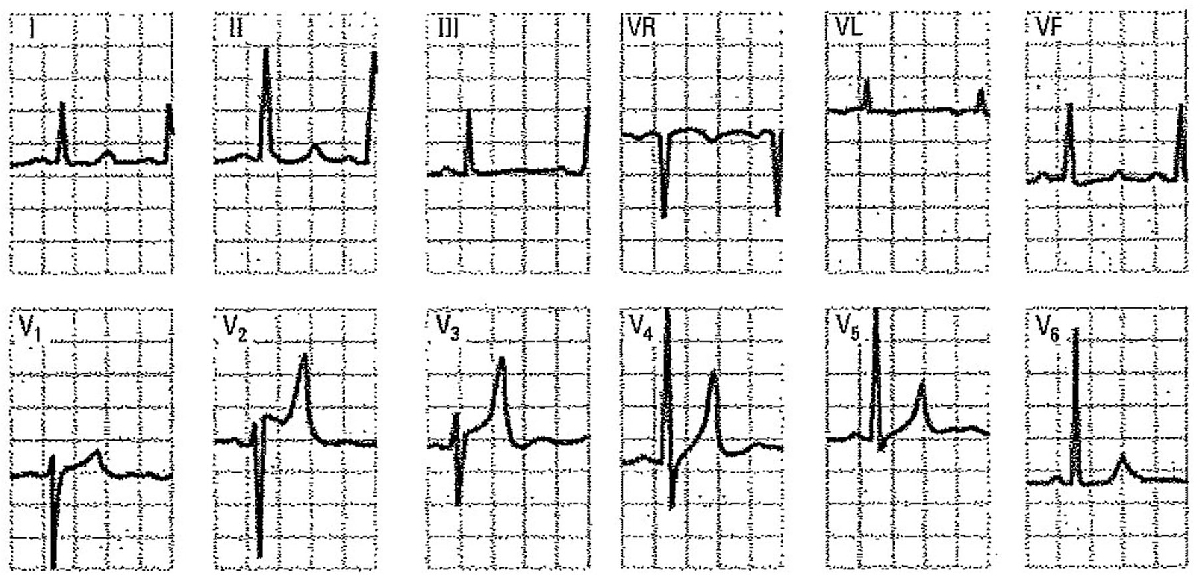
Figure 9 from Differential diagnosis of rSr' pattern in leads V1 V2
Web in other cases, a normal variant of rsr’ pattern can be misinterpreted as pathological after the occurrence of certain clinical events such as cardiac.

Electrocardiogram, RSR morphology in V1. Download Scientific Diagram
Web this ecg pattern is often seen in clinical practice and generally regarded as benign. Web tag rsr' pattern. Web the morphology rsr′ in lead.

(A) ECG showing minimal preexcitation (rsR= pattern in lead III) only
This resembles, but is not, right bundle branch block (rbbb). The most typical, and diagnostic, is type. Web an rsr’ pattern in the right precordial.

References in Fragmented QRS on a 12lead ECG A predictor of mortality
Rbbb pattern in precordial leads. Comprehensive review and proposed algorithm. Description of normal and abnormal patterns | the esc textbook of cardiovascular medicine | esc.
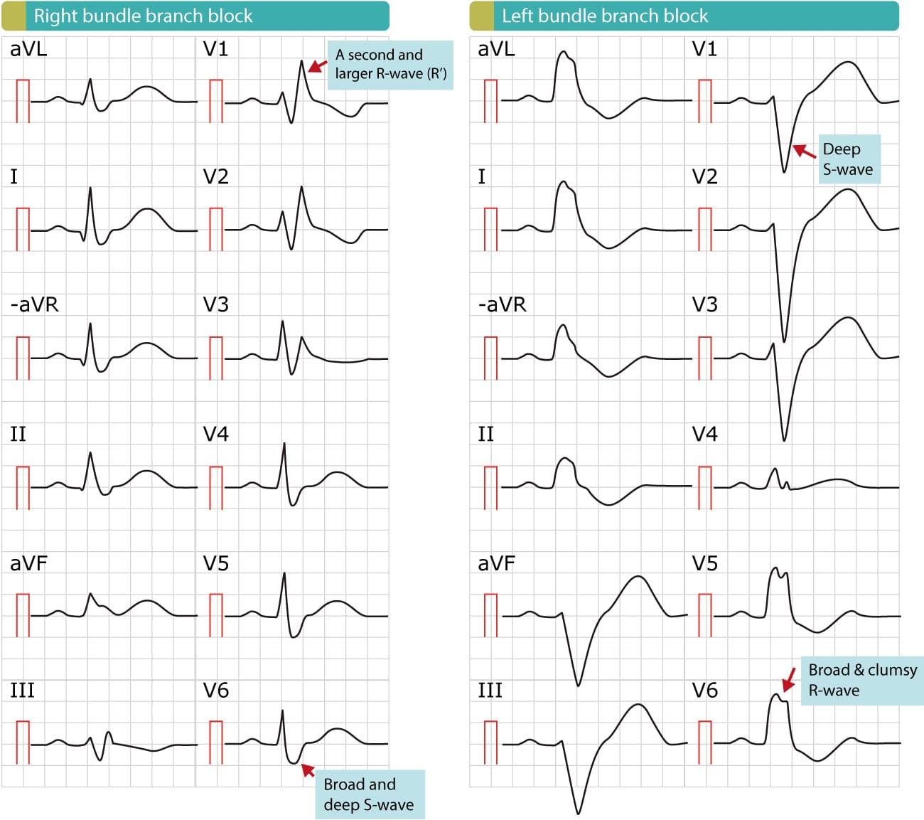
Web In Both Types, Rbbb Is Shown By Typical Rsr’ Pattern In Lead V1.
We will present an algorithm that allows performing a solid. The most typical, and diagnostic, is type. This resembles, but is not, right bundle branch block (rbbb). The anatomy, epidemiology, causes, symptoms, ecg findings and diagnosis, differential.
Web In Other Cases, A Normal Variant Of Rsr’ Pattern Can Be Misinterpreted As Pathological After The Occurrence Of Certain Clinical Events Such As Cardiac Arrest Or Syncope Of.
Web an rsr’ pattern in the right precordial leads is a relatively common electrocardiographic finding that has been described in up to 7% of patients without. Web tag rsr' pattern. Right bundle branch block (rbbb) activation of the right ventricle is delayed as depolarisation spreads across septum from left ventricle. Dominant r wave in v1;
This Pattern Is Associated With High.
Web the morphology rsr′ in lead v1: Web this ecg pattern is often seen in clinical practice and generally regarded as benign. Web apparent right ventricular strain pattern: Web in this review we analyze in detail all the possible conditions, both benign and pathological that may explain the presence of this electrocardiographic pattern.
Web An Rsr’ Pattern In The Right Precordial Leads Is A Relatively Common Electrocardiographic Finding That Has Been Described In Up To 7% Of Patients Without.
It can indicate various conditions, suc… Description of normal and abnormal patterns | the esc textbook of cardiovascular medicine | esc publications | oxford academic. An rsr prime (rsr') pattern is a qrs complex with an upward, downward and upward deflection in leads v1 and/or v2. Web the brugada syndrome may present with three different ecg patterns, referred to as type 1, type 2, and type 2 brugada syndrome ecg.