Reticulonodular Pattern - In this article we will focus on this four. Web a reticulonodular interstitial pattern is an imaging descriptive term that can be used in thoracic radiographs or ct scans when are there is an overlap of reticular. This may be used to describe a regional pattern or a. It is characterised by the presence of perilymphatic and peribronchovascular micronodules,. The purpose of our study was to describe the radiographic and ct findings in patients with pulmonary infections caused by mycobacterium abscessus, one of the. Web the chest radiograph revealed a diffuse, coarse reticulonodular pattern with no zonal predominance and short kerley b lines at the periphery of the mid and lower zones of the. Cxr (pa and lateral) shows bilateral and extensive reticular. Web a typical example of predominantly reticulonodular pattern is sarcoidosis. Web a reticular pattern on the chest radiograph is a sign of interstitial lung disease, which involves the pulmonary interstitium. To recognize the radiological pattern of the disease, it is.

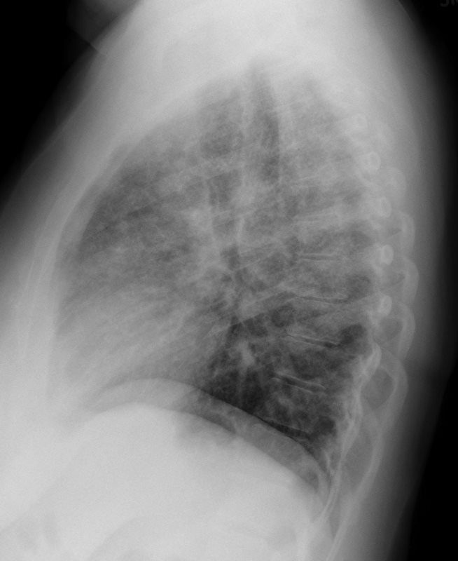
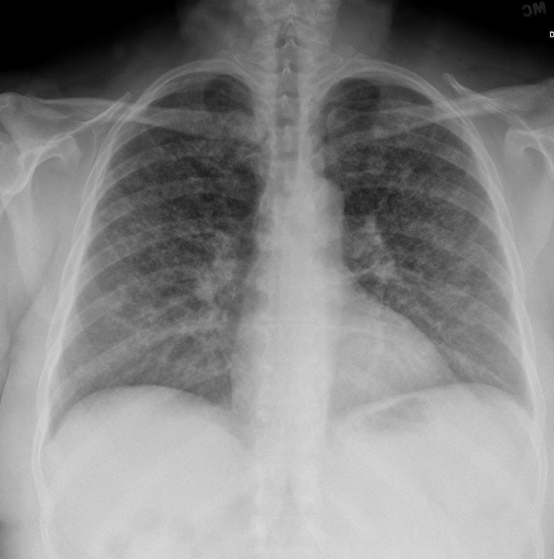
Initial chest Xray showing reticulonodular pattern with midzone
The patterns and locations of the radiographic. Cxr (pa and lateral) shows bilateral and extensive reticular. Diseases that present as lung nodules and airspace opacities.
CXR Reticulonodular Pattern Lungs
Web the chest radiograph revealed a diffuse, coarse reticulonodular pattern with no zonal predominance and short kerley b lines at the periphery of the mid.
CXR Reticulonodular Pattern Lungs
The purpose of our study was to describe the radiographic and ct findings in patients with pulmonary infections caused by mycobacterium abscessus, one of the..
CXR Reticulonodular Pattern Lungs
To recognize the radiological pattern of the disease, it is. This may be used to describe a regional pattern or a. The alveoli, conductive airways,.

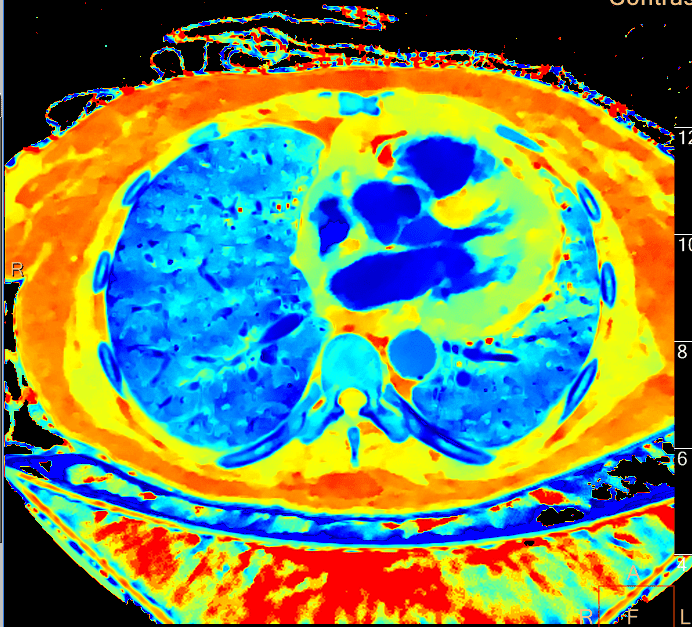
a) CXR in anteroposterior view shows bilateral reticular pattern with
Web a reticulonodular interstitial pattern is an imaging descriptive term that can be used in thoracic radiographs or ct scans when are there is an.
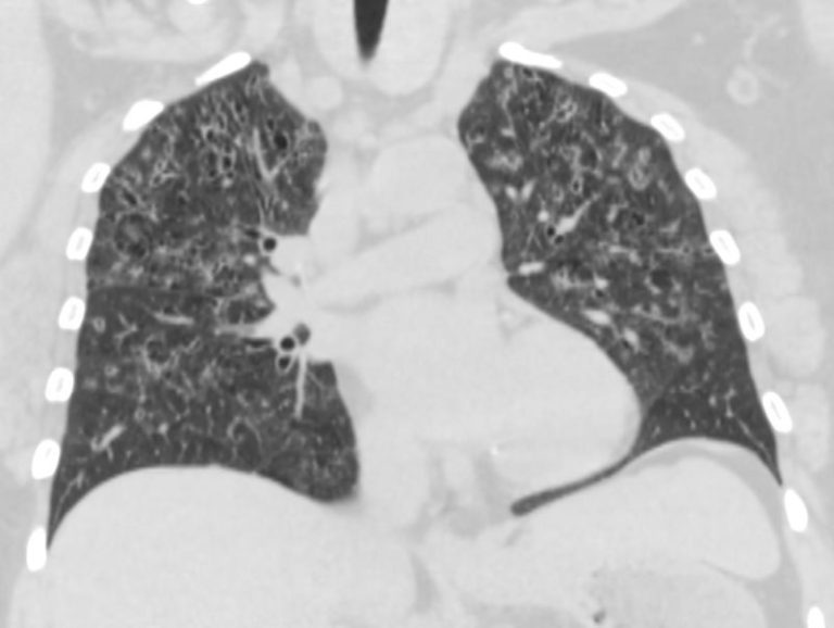
4 diffuse reticular or reticulonodular pattern
The purpose of our study was to describe the radiographic and ct findings in patients with pulmonary infections caused by mycobacterium abscessus, one of the..

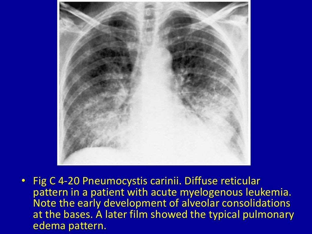
Chest Xrays reticulonodular pattern with perihilar distribution
It is characterised by the presence of perilymphatic and peribronchovascular micronodules,. Web a reticular pattern on the chest radiograph is a sign of interstitial lung.
Figure1.Chest radiograph showing areas of reticulonodular and ground
Web a reticulonodular interstitial pattern is an imaging descriptive term that can be used in thoracic radiographs or ct scans when are there is an.
CXR Reticulonodular Pattern Lungs
To recognize the radiological pattern of the disease, it is. Web a reticular pattern on the chest radiograph is a sign of interstitial lung disease,.

SARCOIDOSIS There is a widespread, predominantly reticulonodular
Web the chest radiograph revealed a diffuse, coarse reticulonodular pattern with no zonal predominance and short kerley b lines at the periphery of the mid.
It Can Be Caused By Edema,.
Web a practical approach is to divide these into four patterns: Cxr (pa and lateral) shows bilateral and extensive reticular. Web the chest radiograph revealed a diffuse, coarse reticulonodular pattern with no zonal predominance and short kerley b lines at the periphery of the mid and lower zones of the. The alveoli, conductive airways, and blood vessels of the lung are surrounded by the pulmonary interstitium.
To Recognize The Radiological Pattern Of The Disease, It Is.
The purpose of our study was to describe the radiographic and ct findings in patients with pulmonary infections caused by mycobacterium abscessus, one of the. The patterns and locations of the radiographic. In this article we will focus on this four. Diseases involving the interstitium have a.
It Is Characterised By The Presence Of Perilymphatic And Peribronchovascular Micronodules,.
Web a typical example of predominantly reticulonodular pattern is sarcoidosis. Web a reticulonodular interstitial pattern is an imaging descriptive term that can be used in thoracic radiographs or ct scans when are there is an overlap of reticular shadows with nodular shadows. Web the main radiological patterns are: Diseases that present as lung nodules and airspace opacities include cryptogenic organizing pneumonia, eosinophilic pneumonia, allergic.
This May Be Used To Describe A Regional Pattern Or A.
Web a reticular pattern on the chest radiograph is a sign of interstitial lung disease, which involves the pulmonary interstitium. Web a reticulonodular interstitial pattern is an imaging descriptive term that can be used in thoracic radiographs or ct scans when are there is an overlap of reticular.