Reticulonodular Pattern On Cxr - A practical approach is to divide these into four patterns: Web reticulation can be subdivided by the size of the intervening pulmonary lucency into fine, medium and coarse. A reticulonodular pattern results from a combination of reticular and nodular opacities. For example, kaposi sarcoma (ks). Diagnosis was confirmed by a transbronchial biopsy. Diseases with a predominantly reticular pattern can be subdivided by the acuteness of their presentation. Interlobular septal thickening is an. The scarring associated with interstitial lung disease eventually affects your ability to breathe and get enough oxygen into your bloodstream. Web pattern recognition should be combined with knowledge of clinical factors in order to generate a limited and meaningful differential diagnosis [table 2]. Make a specific diagnosis of ild when supportive findings are present in the history or on radiologic imaging (e.g., dilated esophagus and ild in scleroderma.

Chest Xrays reticulonodular pattern with perihilar distribution
Web an explanation of alveolar vs. The wbc count was 9.4×10 9 /l with neutrophilia. The scarring associated with interstitial lung disease eventually affects your.

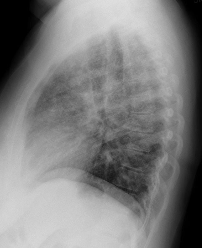
Chest X‑ray PA view showing reticulonodular markings in bilateral lung
Web the chest radiograph revealed a diffuse, coarse reticulonodular pattern with no zonal predominance and short kerley b lines at the periphery of the mid.
CXR Reticulonodular Pattern Lungs
A reticulonodular interstitial pattern is produced by either overlap of reticular shadows or by the presence of reticular shadowing and pulmonary nodules.while this is a.

Interstitial Changes Chest X Ray Medschool vrogue.co
Many people diagnosed with interstitial lung diseases are initially treated with a corticosteroid (prednisone), sometimes in combination with other drugs that suppress the immune system..
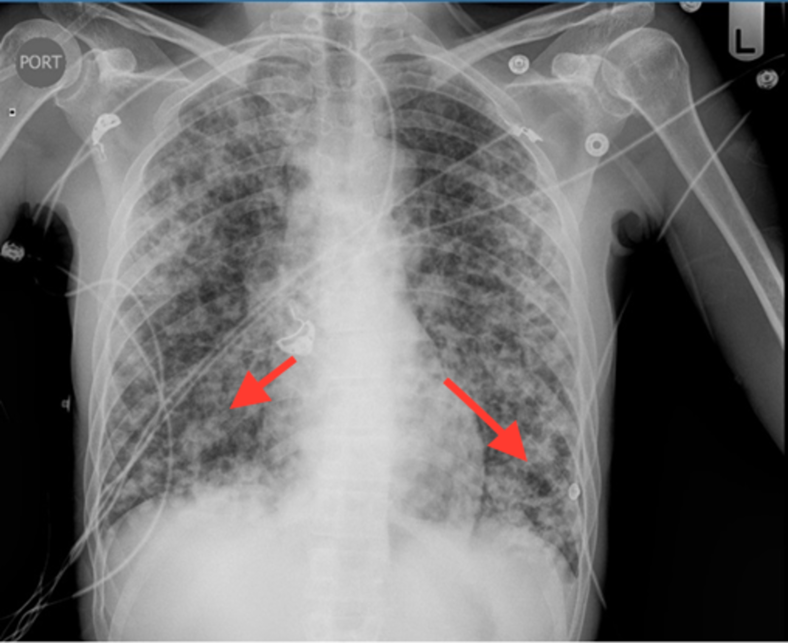
Cureus Recurrent Pneumocystis Pneumonia with Radiographic
Make a specific diagnosis of ild when supportive findings are present in the history or on radiologic imaging (e.g., dilated esophagus and ild in scleroderma..
CXR Reticulonodular Pattern Lungs
Web the chest radiograph revealed a diffuse, coarse reticulonodular pattern with no zonal predominance and short kerley b lines at the periphery of the mid.
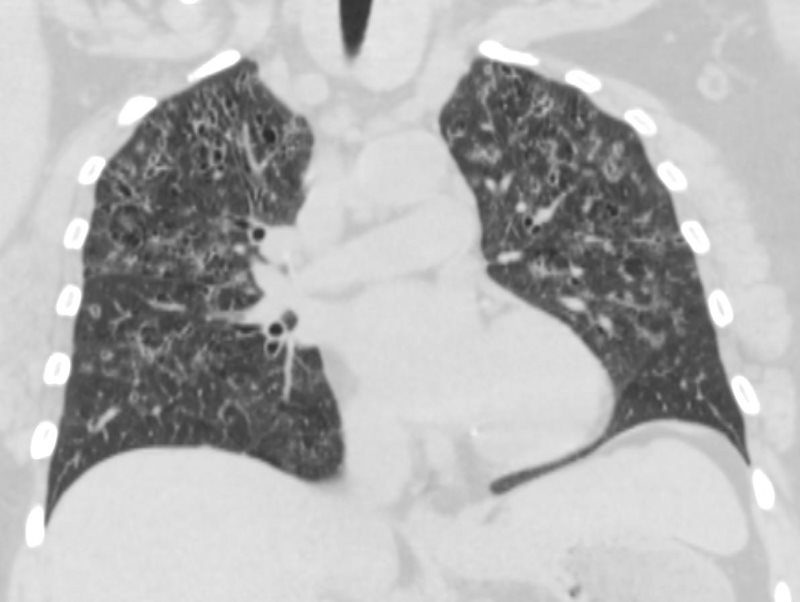
Figure1.Chest radiograph showing areas of reticulonodular and ground
Interlobular septal thickening is an. A reticulonodular pattern results from a combination of reticular and nodular opacities. The scarring associated with interstitial lung disease eventually.

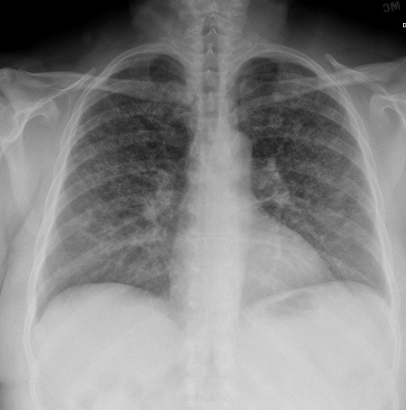
Chest Xray AP view showing reticulonodular infiltrates and
Make a specific diagnosis of ild when supportive findings are present in the history or on radiologic imaging (e.g., dilated esophagus and ild in scleroderma..
CXR Reticulonodular Pattern Lungs
Web pattern recognition should be combined with knowledge of clinical factors in order to generate a limited and meaningful differential diagnosis [table 2]. The wbc.
The Radiology Assistant Chest XRay Lung disease
Web reticulation can be subdivided by the size of the intervening pulmonary lucency into fine, medium and coarse. Web an interstitial lung pattern is a.
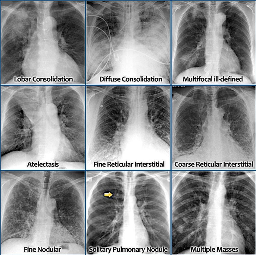
Reticular—Fine Or Coarse Linear Shadows;
Reticular opacities seen on hrct in patients with diffuse lung disease can indicate lung infiltration with interstitial thickening or fibrosis. Web list and identify on a chest radiograph and computed tomographic (ct) scan the four patterns of interstitial lung disease (ild): Acute, not a common pattern. For example, kaposi sarcoma (ks).
A Reticulonodular Interstitial Pattern Is Produced By Either Overlap Of Reticular Shadows Or By The Presence Of Reticular Shadowing And Pulmonary Nodules.while This Is A Relatively Common Appearance On A Chest Radiograph, Very Few Diseases Are Confirmed To Show This Pattern Pathologically.examples Include:
Associated with signs of volume loss; Many diverse pathological processes can cause diffuse lung disease. When infiltrates are present, the particular pattern is of limited value for differentiating among cardiogenic pulmonary edema, noncardiogenic, pulmonary edema, hemorrhage, atelectasis, and pneumonia. Web the chest radiograph revealed a diffuse, coarse reticulonodular pattern with no zonal predominance and short kerley b lines at the periphery of the mid and lower zones of the left lung.
These Are Interlobular Septal Thickening, Honeycombing, And Irregular Reticulation.
Can sometimes give a fine reticulonodular pattern 3. Make a specific diagnosis of ild when supportive findings are present in the history or on radiologic imaging (e.g., dilated esophagus and ild in scleroderma. Web atypical pneumonia is a term used inconsistently through time and in different parts of the world. Nodular—small (2 to 3 mm), medium, large, or masses (>3 cm) 3.
The Scarring Associated With Interstitial Lung Disease Eventually Affects Your Ability To Breathe And Get Enough Oxygen Into Your Bloodstream.
Diseases with a predominantly reticular pattern can be subdivided by the acuteness of their presentation. Web based on currently available, scientific evidence, however, your doctor may recommend: Imaging findings include a bronchopneumonia pattern with centrilobular nodules, bronchial wall thickening, ggo, and patchy consolidation, which later leads to lobar distribution. Many people diagnosed with interstitial lung diseases are initially treated with a corticosteroid (prednisone), sometimes in combination with other drugs that suppress the immune system.