Reticular Pattern On Chest X Ray - Reticular pattern and bronchiectasis, involving predominantly lower lobes and costophrenic angles, are generally recognised. The prematurity of the imaging test and the absence of pulmonary disease at the time of presentation; Although distinction between these abnormalities often is. Web detecting diffuse lung infiltrates on chest radiography is a common clinical problem. Unfortunately, imaging findings could be misdiagnosed in the early stage of disease. This may be used to describe a regional pattern or a diffuse pattern throughout the lungs. Unfortunately, imaging findings could be misdiagnosed in the early stage of disease. Here a cxr with a reticular pattern at the lung bases. A reticular pattern is characterized by innumerable interlacing line shadows that suggest a mesh ( fig. Web reticular patterns represent interstitial lung disease.

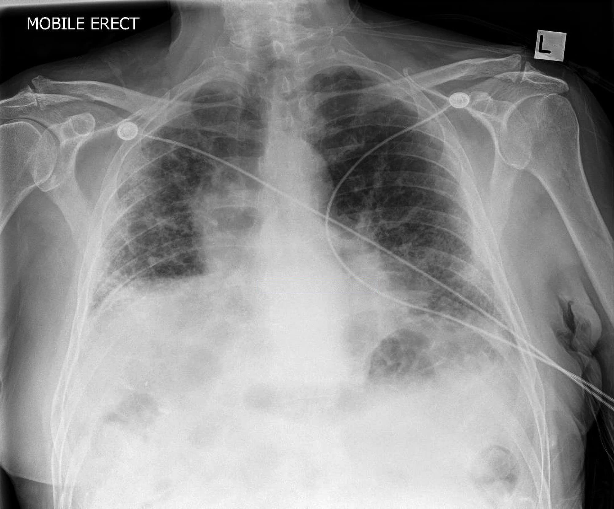
a) CXR in anteroposterior view shows bilateral reticular pattern with
Web coarse reticular pattern. Also seen when pneumonia or pulmonary edema occurs in patients with underlying emphysema; Three principal patterns of reticulation may be seen..

Chest Xray multiple bilateral opacities and reticular pattern in both
Web what is an interstitial lung pattern? In many cases you can suspect uip on the cxr. A common radiographic pattern that encompasses the same.

Chest Xray at the first visit showing bilateral reticular opacity
Three principal patterns of reticulation may be seen. Web what is an interstitial lung pattern? Reticulation can be subdivided by the size of the intervening..

Chest Xray showing diffuse reticular changes. Download Scientific
A reticulonodular interstitial pattern is an imaging descriptive term that can be used in thoracic radiographs or ct scans when are there is an overlap.
Reticular Chest X Ray
Interlobular septal thickening is an. Web detecting diffuse lung infiltrates on chest radiography is a common clinical problem. This is the lung tissue between the.

Interstitial pneumonias An acute reticular pattern GrepMed
Three principal patterns of reticulation may be seen. This is the lung tissue between the spaces that are filled with air in the lung. Web.

Chest X Ray Reticular Pattern
This says nothing of the cause or diagnosis. Although distinction between these abnormalities often is. Unfortunately, imaging findings could be misdiagnosed in the early stage.
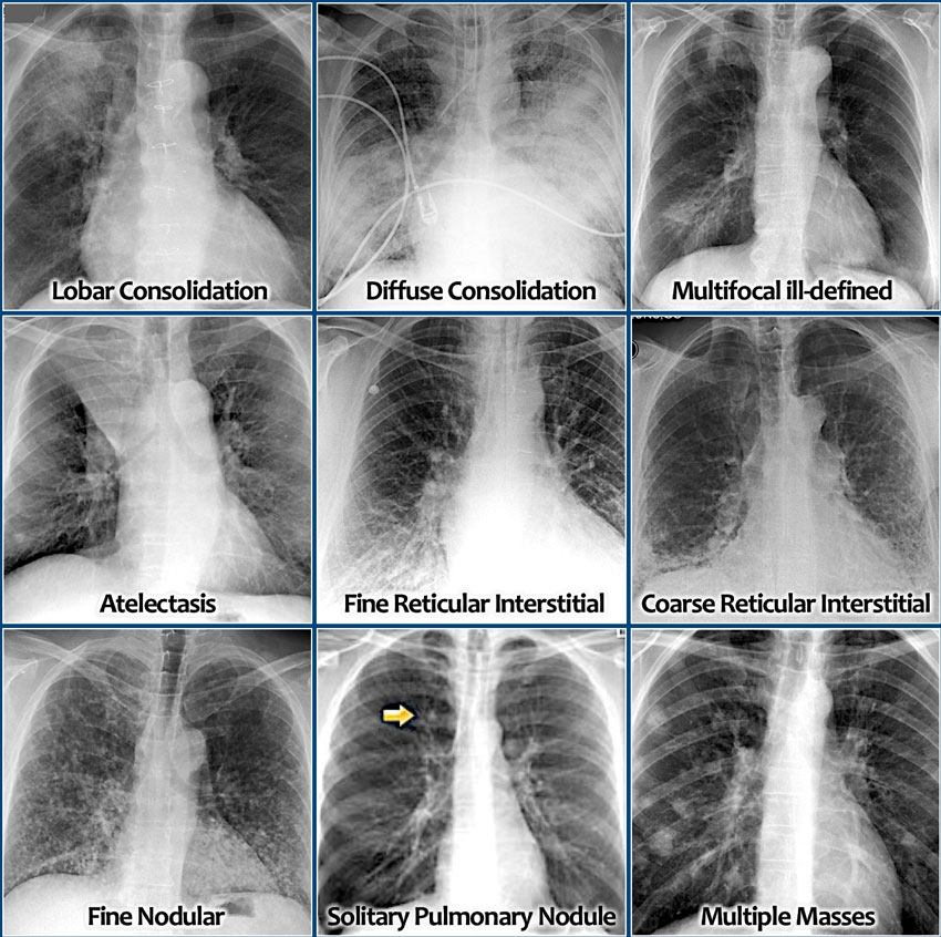
The Radiology Assistant Chest XRay Lung disease
Linear, reticular, reticulonodular, and nodular. Interlobular septal thickening is an. Web coarse reticular pattern. A reticular pattern is characterized by innumerable interlacing line shadows that.

Chest radiography showing increased reticular markings and areas of
A common radiographic pattern that encompasses the same disorders as reticular patterns; Reticulation can be subdivided by the size of the intervening. Unfortunately, imaging findings.

Reticular Chest X Ray
Reticular pattern and bronchiectasis, involving predominantly lower lobes and costophrenic angles, are generally recognised. A reticular pattern is characterized by innumerable interlacing line shadows that.
Many Diverse Pathological Processes Can Cause Diffuse Lung Disease.
Also seen when pneumonia or pulmonary edema occurs in patients with underlying emphysema; This says nothing of the cause or diagnosis. This may be used to describe a regional pattern or a diffuse pattern throughout the lungs. Unfortunately, imaging findings could be misdiagnosed in the early stage of disease.
Make A Specific Diagnosis Of Ild When Supportive Findings Are Present In The History Or On Radiologic Imaging (E.g., Dilated Esophagus And Ild In Scleroderma.
Plain chest radiography remains the first diagnostic approach to diffuse infiltrative lung disease but has limited diagnostic sensitivity and specificity. A hrct is needed to confirm the diagnosis by demonstrating honeycombing. Interlobular septal thickening is an. Reticular pattern and bronchiectasis, involving predominantly lower lobes and costophrenic angles, are generally recognised.
Although Distinction Between These Abnormalities Often Is.
This finding means that there is abnormality of the support tissues of the lung. Three principal patterns of reticulation may be seen. A reticulonodular interstitial pattern is produced by either overlap of reticular shadows or by the presence of reticular shadowing and pulmonary nodules.while this is a relatively common appearance on a chest radiograph, very few diseases are confirmed to show this pattern pathologically.examples include: Here a cxr with a reticular pattern at the lung bases.
A Reticular Pattern Is Characterized By Innumerable Interlacing Line Shadows That Suggest A Mesh ( Fig.
Web coarse reticular pattern. Web an interstitial lung pattern is a regular descriptive term used when reporting a plain chest radiograph. This is the lung tissue between the spaces that are filled with air in the lung. Web what is an interstitial lung pattern?