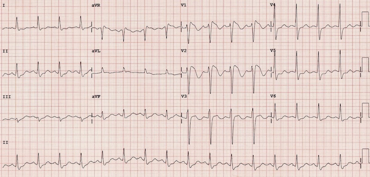
Qr Pattern In V1 - Many ecg signs are more frequent in patients with pulmonary embolism. Comprehensive review and proposed algorithm. Web qrs morphologies in v1 and v6 during left bundle branch area pacing: Web an rsr’ pattern in the right precordial leads is a relatively common electrocardiographic finding that has been described in up to 7% of patients without apparent heart disease. Web clockwise rotation of the qrs vector in the precordial leads (clockrot) defined as r = s in v4, v5 or v6. Web as v1 is a unipolar lead, structures closer to the chest wall show a lbbb pattern with a qs complex, while more posterior structures show a progressive increase in the initial r wave amplitude through a right bundle branch block (rbbb) pattern. Web presence of a qr complex in lead v1 had a 96% specificity but r:s ratio, voltage criteria and rsr' incomplete right bundle branch block pattern had intermediate specificities of 66%, 66% and 52%, respectively. 4 if the qrs is wide, the presence of an r’ in leads v 1 ‐v 2 usually is in the context of a complete right bundle branch block (rbbb), but other causes have. Web rsr′ or qr pattern in v1 suggests right ventricular conduction delay. Web paced qrs morphological pattern in lead v1 was most commonly qr pattern followed by qr pattern.
rsr or qr pattern in v1 performingartsphotographybybateman
Web from a vectorcardiographic perspective, this pattern suggests a posterior orientation of initial qrs forces, an important cause of which could be myocardial infarction (mi).

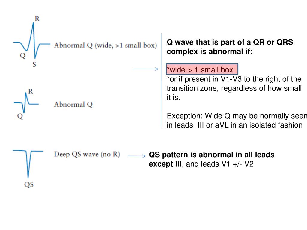
A Qr pattern in V1 indicates that the lead tip has reached the left
R in v1 + s in v5 (or v6) 10 mm; 1 septal, or mid‐septal infarction is an ecg diagnosis that has been used. When.
PPT ECG 1 PowerPoint Presentation, free download ID6591352
Web with complete bundle branch blocks, the qrs interval is classically stated to be greater than or equal to 120 ms (0.12 s) in duration.

Rsr Or Qr Pattern In V1 / The Rsr Pattern In Leads V1 V2 Algorithm And
Many ecg signs are more frequent in patients with pulmonary embolism. R in v5 or v6 < 5 mm ; Web an rsr’ pattern v1.

rsr or qr pattern in v1 makenafefge
1 septal, or mid‐septal infarction is an ecg diagnosis that has been used. Web the qrs interval of the right ventricle is marked positive. With.

Understanding QR Codes Mechanism, Pros, and Terminology
Web rsr′ or qr pattern in v1 suggests right ventricular conduction delay. Many ecg signs are more frequent in patients with pulmonary embolism. R/s ratio.

Rsr Or Qr Pattern In V1 / File De Brugada Ecg Characteristics
Electrocardiography (ecg) in patients with pulmonary embolism may show several abnormalities related to right ventricular strain. Web clockwise rotation of the qrs vector in the.

PPT Normal ECG Rate and Rhythm PowerPoint Presentation, free
Web in six patients whose vt arose from the middle part of the amc, we demonstrated a special (‘rebound’) transition pattern, with which equal r.

rsr or qr pattern in v1 performingartsphotographybybateman
As a library, nlm provides access to scientific literature. Comprehensive review and proposed algorithm. Web qr in v 1 reflects right ventricle dilation and interventricular.

Right Bundle Branch Block (RBBB) • LITFL • ECG Library Diagnosis
R in v1 + s in v5 (or v6) 10 mm; Web presence of a qr complex in lead v1 had a 96% specificity but.
R In V1 + S In V5 (Or V6) 10 Mm;
4 if the qrs is wide, the presence of an r’ in leads v 1 ‐v 2 usually is in the context of a complete right bundle branch block (rbbb), but other causes have. With incomplete blocks, the qrs interval is defined between 100 (or 110 by computer) and 120 ms (0.10 [or 0.11 by computer] to 0.12 s). Web any one of the following in lead v1: Web as v1 is a unipolar lead, structures closer to the chest wall show a lbbb pattern with a qs complex, while more posterior structures show a progressive increase in the initial r wave amplitude through a right bundle branch block (rbbb) pattern.
Web From A Vectorcardiographic Perspective, This Pattern Suggests A Posterior Orientation Of Initial Qrs Forces, An Important Cause Of Which Could Be Myocardial Infarction (Mi) Underlying These Leads.
When we compare complete and incomplete right bundle branch blocks, the duration of qrs varies. Rsr’ pattern in v1, with (appropriate) discordant t wave changes. Web ivas originating from amc exhibit an lbbb qrs pattern and a qr pattern in lead v1 resulting from its unique location which leads to primordial septal activation followed by rapid lateral to medial basal lv activation (dixit et al., 2005). R/s ratio > 1 and negative t wave ;
Among The Ecg Signs Seen In Patients With Acute Pulmonary Embolism, Qr In V (1)Is Closely Related To The Presence Of Right Ventricular Dysfunction, And Is An Independent Predictor Of Adverse Clinical Outcome.
Comprehensive review and proposed algorithm. The criteria for the diagnosis of qr in v1 were the presence of a prominent q wave of ≥0.2 mv and a ventricular depolarisation v1</strong> ≥ 0.1 mv. Web the qrs interval of the right ventricle is marked positive. As a library, nlm provides access to scientific literature.
Web Qrs Morphologies In V1 And V6 During Left Bundle Branch Area Pacing:
Electrocardiography (ecg) in patients with pulmonary embolism may show several abnormalities related to right ventricular strain. 2 , 3 , 4 however, there is much evidence to indicate that. Web an rsr’ pattern in the right precordial leads is a relatively common electrocardiographic finding that has been described in up to 7% of patients without apparent heart disease. Web rsr′ or qr pattern in v1 suggests right ventricular conduction delay.