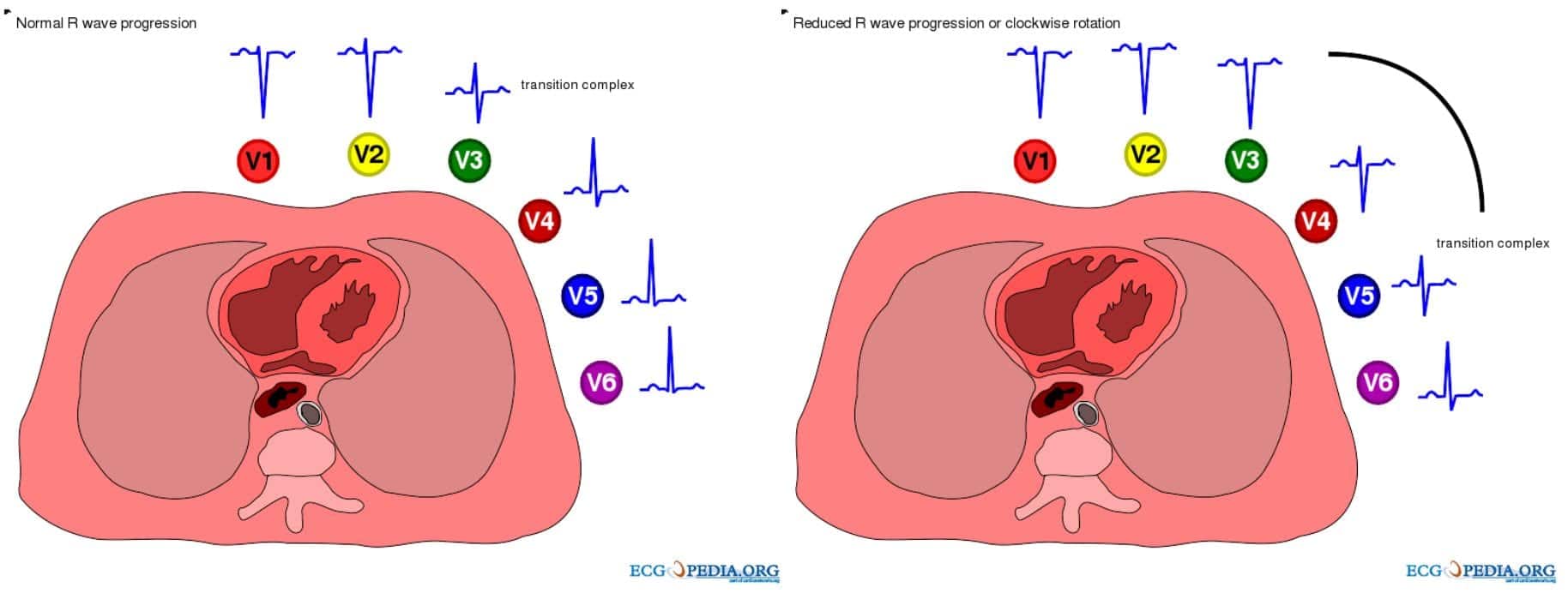
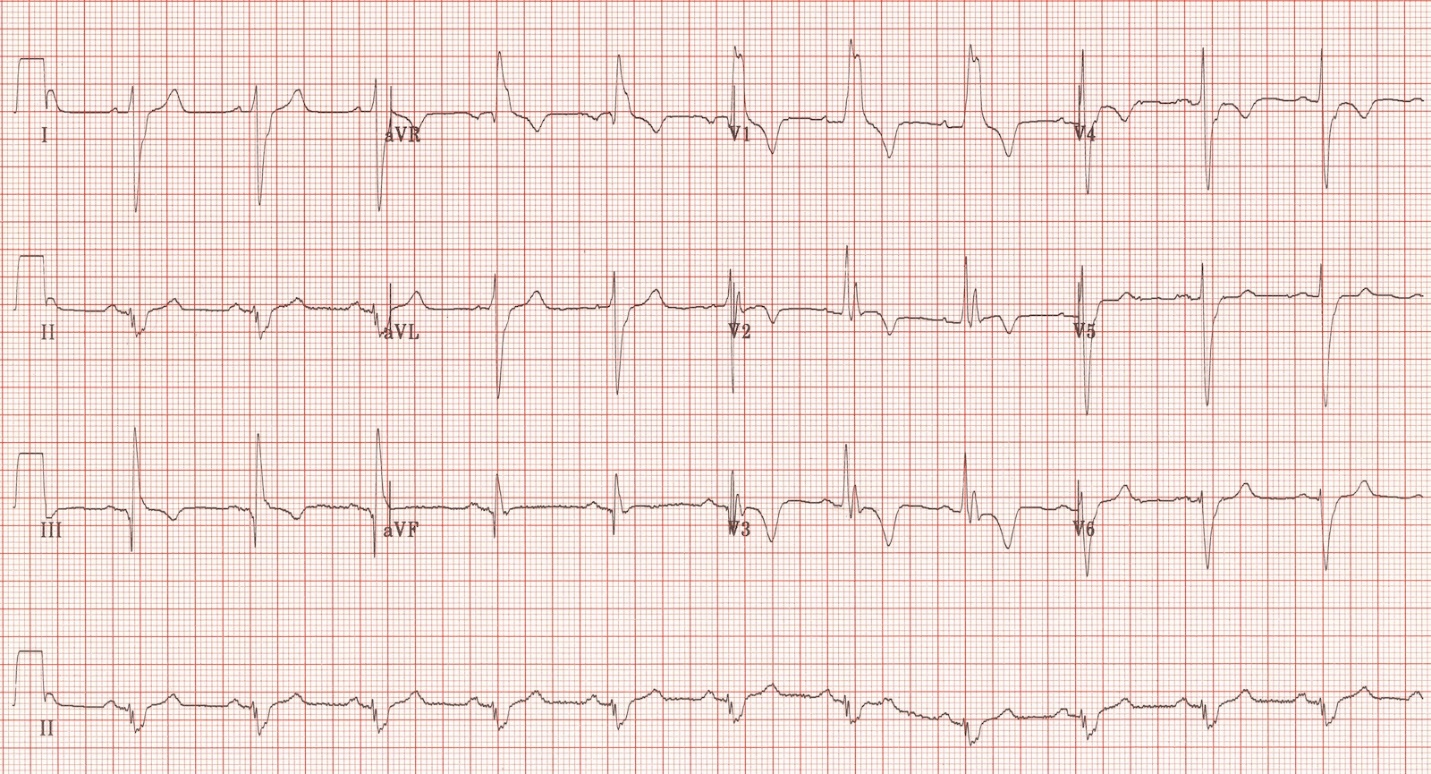
Pulmonary Disease Pattern On Ekg - Web electrocardiographic (ecg) abnormalities associated with chronic obstructive pulmonary disease (copd) include right atrial enlargement, right ventricular hypertrophy, right bundle branch block (rbbb), marked clockwise rotation with poor r‐wave progression, low voltage in the limb leads, a s 1 s 2 s 3 pattern, a qs. Sinus tachycardia or other types of arrhythmias such as atrial flutter or atrial fibrillation. Web left atrial enlargement is also referred to as p mitrale, and right atrial enlargement is often referred to as p pulmonale. Web ecg demonstrates many of the features of chronic pulmonary disease: Web specific electrocardiographic abnormalities and cardiac arrhythmias are prevalent in chronic obstructive pulmonary disease. Pulmonary embolism on the ekg: •right axis deviation of the p waves. Web sinus tachycardia is the most common ecg finding in pulmonary embolism. Web the principal electrocardiogram (ecg) changes associated with ventricular hypertrophy are increases in qrs amplitude and duration, changes in instantaneous and mean qrs vectors, abnormalities in the st segment and t waves, and abnormalities in the p wave. •right axis deviation or vertical axis of the qrs complex.

Pulmonary embolism and S1Q3 pattern Cardiocases
Web patients with chronic obstructive pulmonary disease (copd) often have abnormal ecgs. Our aim was to separate the effects on ecg by airway obstruction, emphysema.

Pulmonary Embolism (PE) Causes, symptoms, diagnosis, treatment
Patients presenting with chest pain, these ekg patterns, and troponin elevation are often misdiagnosed with mi. Right bundle branch block, s1q3t3 pattern. The underlying pathophysiology.

S1Q3T3 EKG Classic Pattern in Pulmonary Embolism (Example). Pulmonary
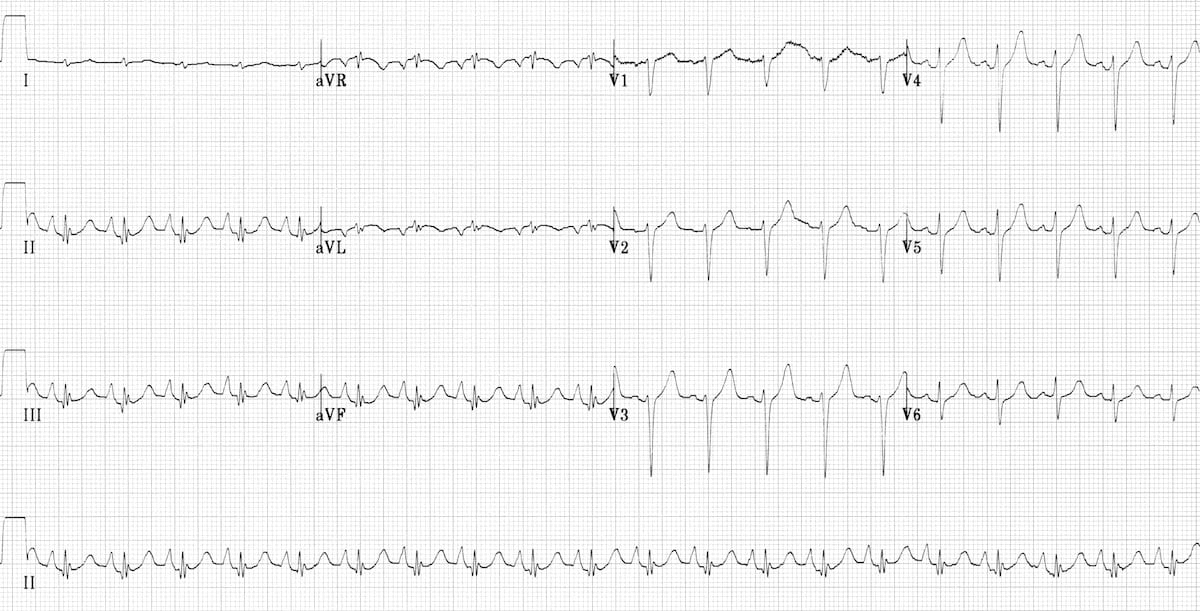
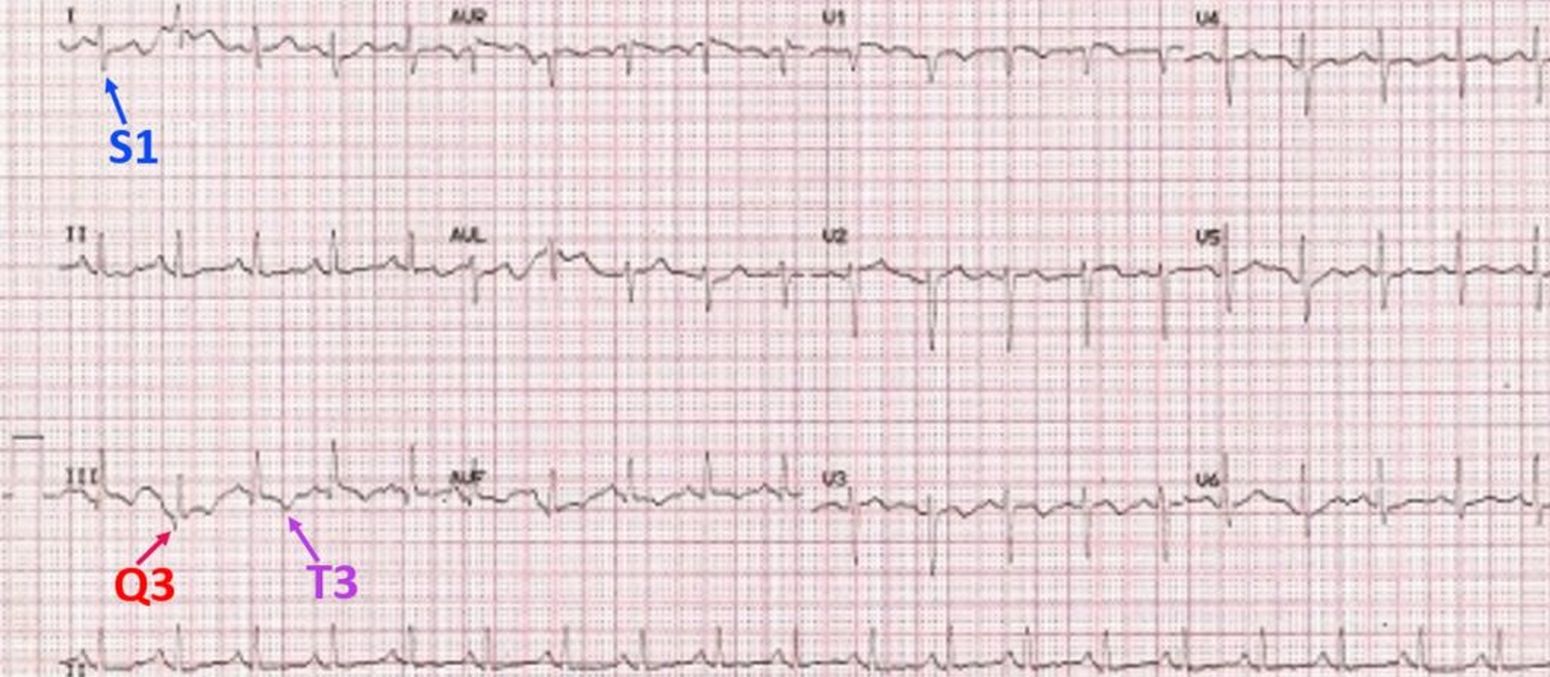
Ecg abnormalities are common in patients with pulmonary embolism, with the most frequent being sinus tachycardia, right ventricular strain, and the classic s1q3t3 pattern. Objective.
ECG in Chronic Obstructive Pulmonary Disease • LITFL • ECG Library
Web electrocardiography (ecg) is a useful adjunct to other pulmonary tests because it provides information about the right side of the heart and therefore pulmonary.
pulmonary disease pattern ecg Hình ảnh có liên quan Diseases Club
Our aim was to separate the effects on ecg by airway obstruction, emphysema and right ventricular (rv) afterload in patients with copd. Inspite of normal.

S1Q3T3 on ECG in a patient with Acute Pulmonary Embolism GrepMed
Web this post describes two ekg patterns of pe which mimic mi. Ecg abnormalities are common in patients with pulmonary embolism, with the most frequent.
ECG in Chronic Obstructive Pulmonary Disease • LITFL • ECG Library
•right axis deviation or vertical axis of the qrs complex. And decreased progression of r waves in precordial leads. These abnormalities have an impact on.

The ECG's of Pulmonary Embolism Resus
The underlying pathophysiology is complex. Autonomic, electrical, and structural changes play a critical role. Web left atrial enlargement is also referred to as p mitrale,.
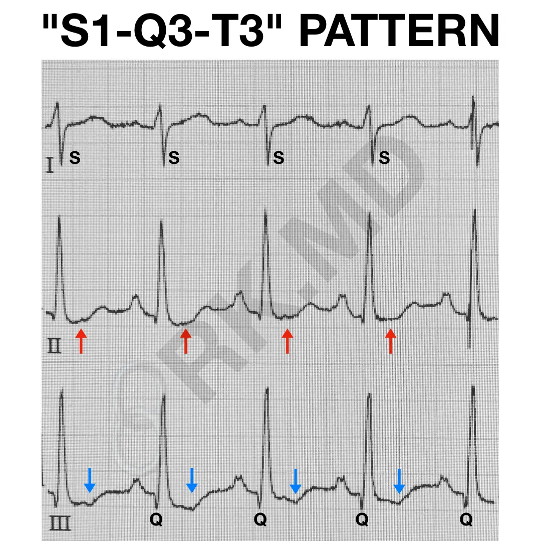
S1Q3T3 EKG Pattern RK.MD
Our aim was to separate the effects on ecg by airway obstruction, emphysema and right ventricular (rv) afterload in patients with copd. Inspite of normal.
ECG Changes in Pulmonary Embolism New Health Advisor
Electrocardiography, pulmonary hypertension, prognosis, pulmonary. Ecg findings often suggest right ventricular pressure overload or strain. Web electrocardiography (ecg) is a useful adjunct to other pulmonary.
Our Aim Was To Separate The Effects On Ecg By Airway Obstruction, Emphysema And Right Ventricular (Rv) Afterload In Patients With Copd.
Electrocardiography, pulmonary hypertension, prognosis, pulmonary. Our aim was to separate the effects on ecg by airway obstruction, emphysema and right ventricular (rv) afterload in patients with copd. Tobacco smoke and air pollution. And decreased progression of r waves in precordial leads.
Web This Post Describes Two Ekg Patterns Of Pe Which Mimic Mi.
•right axis deviation of the p waves. Web ekg changes suggestive of pulmonary embolism. Web the ecg changes associated with acute pulmonary embolism may be seen in any condition that causes acute pulmonary hypertension, including hypoxia causing pulmonary hypoxic vasoconstriction. Ecg findings often suggest right ventricular pressure overload or strain.
Copd Particularly Chronic Bronchitis Was The Commonest Respiratory Problem Next To Pulmonary Tuberculosis.
Web the following ecg signs reflecting ccp were collected: Ecg abnormalities are common in patients with pulmonary embolism, with the most frequent being sinus tachycardia, right ventricular strain, and the classic s1q3t3 pattern. (see also electrocardiography in cardiovascular disorders.) These abnormalities have an impact on cardiovascular prognosis of these patients.
Aka Cord (Respiratory) Or Coad (Airways) Environmental Factors:
Web specific electrocardiographic abnormalities and cardiac arrhythmias are prevalent in chronic obstructive pulmonary disease. Web electrocardiography (ecg) is a useful adjunct to other pulmonary tests because it provides information about the right side of the heart and therefore pulmonary disorders such as chronic pulmonary hypertension and pulmonary embolism. •right axis deviation or vertical axis of the qrs complex. Web left atrial enlargement is also referred to as p mitrale, and right atrial enlargement is often referred to as p pulmonale.