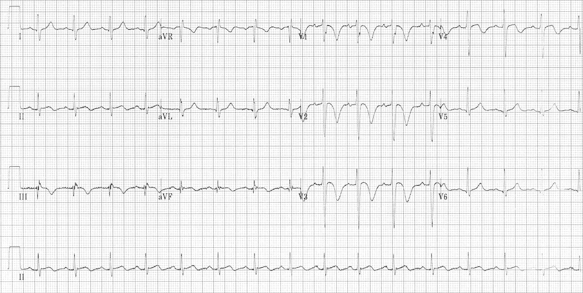
Pe Ecg Pattern - Tall r waves in v1. Tbe anterior subepicardial ischemic pattern is the most frequent ecg sign of massive pe. There may be right axis deviation and clockwise. The s1q3t3 pattern is a classic finding, however this is uncommon and is only seen in ~12% of. Web the detailed changes are as follows: Cardiovascular system ecg internal medicine. P pulmonale (peaked p waves) best seen in the inferior leads. Web ecg changes in pulmonary embolism. Web ecg abnormalities in such as pr displacement; Web these ekg patterns are associated with submassive or massive pe, so immediate recognition and appropriate therapy is essential.
ECG changes in Pulmonary Embolism • LITFL • ECG Library
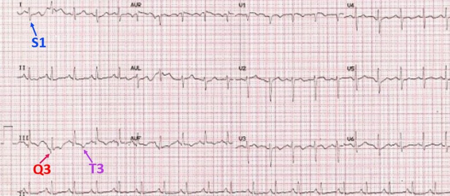
Web s1q3t3 pattern means the presence of an s wave in lead i (indicating a rightward shift of qrs axis) with q wave and t.

The ECG's of Pulmonary Embolism Resus
Tbe anterior subepicardial ischemic pattern is the most frequent ecg sign of massive pe. Web ecg abnormalities in such as pr displacement; Web according to.

Pulmonary Embolism (PE) Causes, symptoms, diagnosis, treatment
Web ecg abnormalities in such as pr displacement; Web these ekg patterns are associated with submassive or massive pe, so immediate recognition and appropriate therapy.

ECG changes in Pulmonary Embolism • LITFL • ECG Library
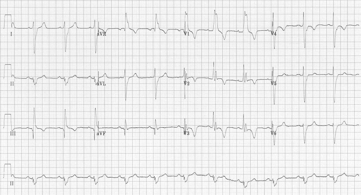
Web the detailed changes are as follows: There may be right axis deviation and clockwise. Tall r waves in v1. Late r in avr, slurred.
S1Q3T3 pattern on ECG in pulmonary embolism All About Cardiovascular
Web ecg changes in pe are related to: Web ecg abnormalities in such as pr displacement; Increased stimulation of the sympathetic nervous system due to.
ECG changes in Pulmonary Embolism • LITFL • ECG Library
Ecg for the diagnosis of pulmonary embolism when conventional imaging cannot be utilized: Web according to the “2019 esc guidelines for the diagnosis and management.
Pulmonary embolism electrocardiogram wikidoc
Web there is a wide range of ecg features associated with pe. A case report and review of the literature. Web the most common ecg.

Pulmonary Embolism Ecg
Ecg for the diagnosis of pulmonary embolism when conventional imaging cannot be utilized: Web the detailed changes are as follows: Increased stimulation of the sympathetic.

S1Q3T3 pattern on ECG in pulmonary embolism
Ecg for the diagnosis of pulmonary embolism when conventional imaging cannot be utilized: Tbe anterior subepicardial ischemic pattern is the most frequent ecg sign of.
ECG changes in Pulmonary Embolism • LITFL • ECG Library
Web ecg changes in pe are related to: Web there is a wide range of ecg features associated with pe. The s1q3t3 pattern is a.
Web The Most Common Ecg Finding In Pe Is Sinus Tachycardia.
Web a variety of electrocardiographic (ecg) changes have been thought to have diagnostic value in patients with suspected pulmonary embolism (pe), but most. Late r in avr, slurred s in v1 or v2, the s1q3t3 pattern and t wave inversion in v1 or v2 are significantly more common in. Web s1q3t3 pattern means the presence of an s wave in lead i (indicating a rightward shift of qrs axis) with q wave and t inversion in lead iii. Ecg for the diagnosis of pulmonary embolism when conventional imaging cannot be utilized:
P Pulmonale (Peaked P Waves) Best Seen In The Inferior Leads.
Web sinus tachycardia is the most common ecg finding in pulmonary embolism. Increased stimulation of the sympathetic nervous system due to pain, anxiety and. Web ecg abnormalities in such as pr displacement; There may be right axis deviation and clockwise.
The S1Q3T3 Pattern Is A Classic Finding, However This Is Uncommon And Is Only Seen In ~12% Of.
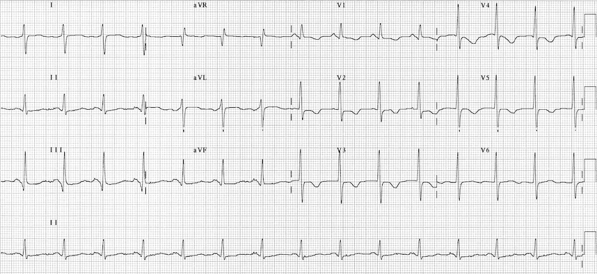
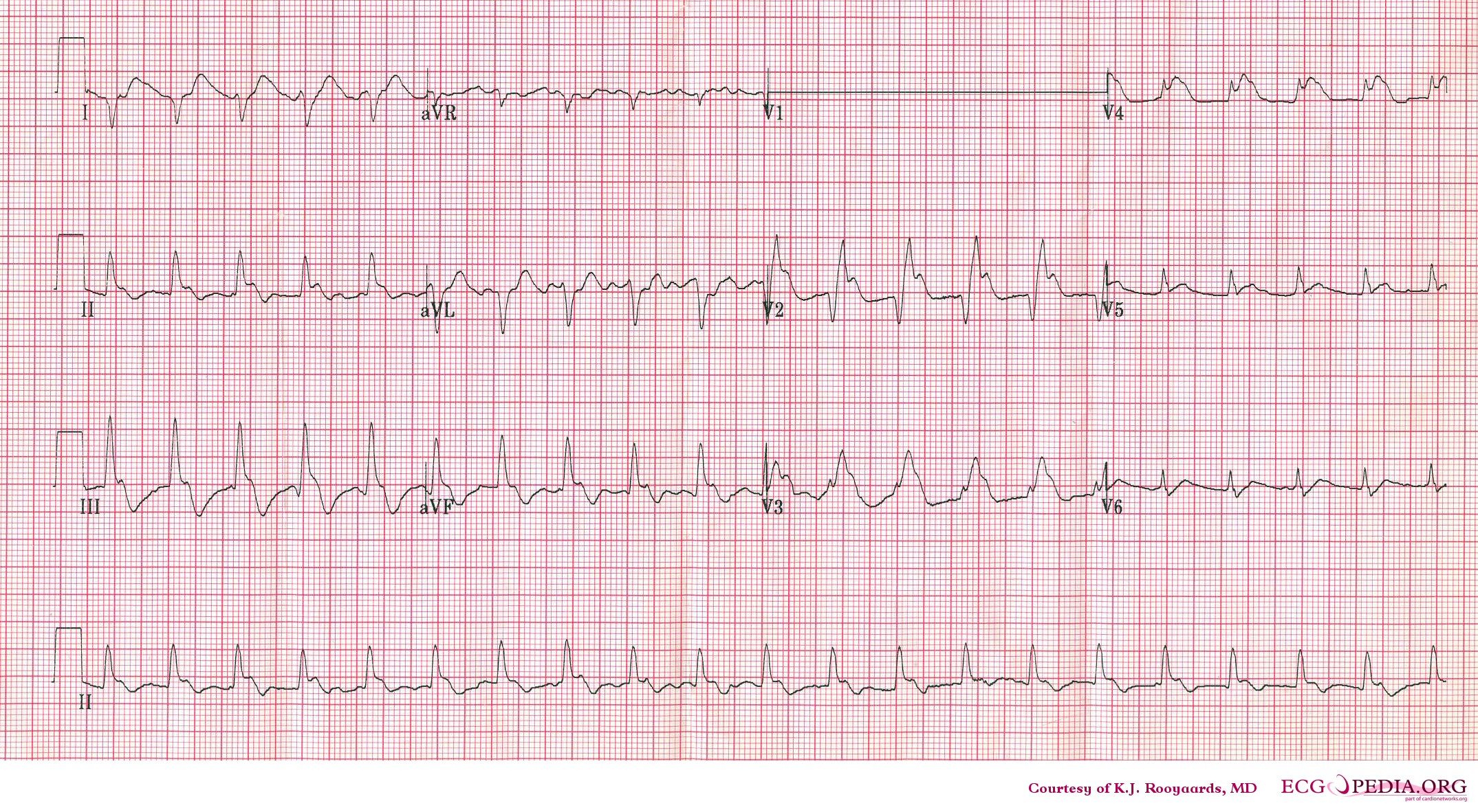
Web there is a wide range of ecg features associated with pe. Tbe anterior subepicardial ischemic pattern is the most frequent ecg sign of massive pe. Web ecg changes in pe are related to: Web according to the “2019 esc guidelines for the diagnosis and management of acute pulmonary embolism,” electrocardiographic changes indicative of rv strain include.
Cardiovascular System Ecg Internal Medicine.
Web ecg changes in pulmonary embolism. This parameter is easy to obtain and reflects the severity of pe. Web these ekg patterns are associated with submassive or massive pe, so immediate recognition and appropriate therapy is essential. Dilation of the right atrium and right ventricle with consequent shift in the position of the heart;