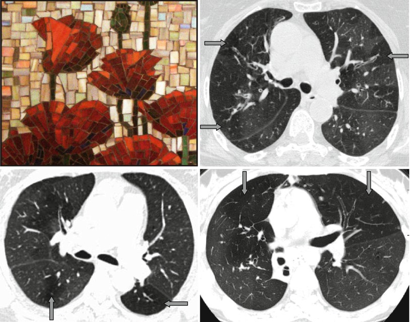
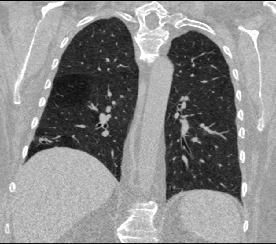
Mosaic Pattern In Lungs - Diseases that primarily affect the small vessels of the lung are difficult to diagnose. To recognize the radiological pattern of the disease, it is. Web by definition, mosaic attenuation is a ct pattern in which areas of differing attenuation are found diffusely distributed throughout the lung parenchyma. Web notably, mosaic attenuation, characterized by patchy areas of differing lung density, may serve as a distinctive marker of cvd such as rheumatoid arthritis 7 or sjögren`s. If vessels in hypoattenuated regions of the lung are smaller than in the other regions, the pattern is due to mosaic perfusion (i.e. Web ct mosaic pattern of lung attenuation: Web both lungs demonstrate multiple regions of mosaic attenuation, most prominent in both lower lobes. Mosaic pattern refers to areas of variable lung attenuation seen on computed tomography (ct) of the chest. Web mosaic lung attenuation 1. This is associated with enlargement of the central pulmonary arteries, with.
mosaic attenuation pattern in lung pacs
This is associated with enlargement of the central pulmonary arteries, with. If vessels in hypoattenuated regions of the lung are smaller than in the other.

Mosaic Attenuation Etiology, Methods of Differentiation, and Pitfalls
If vessels in hypoattenuated regions of the lung are smaller than in the other regions, the pattern is due to mosaic perfusion (i.e. This article.

Mosaic attenuation pattern, CT, Terms YouTube
To recognize the radiological pattern of the disease, it is. Diseases that primarily affect the small vessels of the lung are difficult to diagnose. Web.

Mosaic Attenuation Etiology, Methods of Differentiation, and Pitfalls
To recognize the radiological pattern of the disease, it is. Web by definition, mosaic attenuation is a ct pattern in which areas of differing attenuation.
Perfusion or Mosaic Lung Sign Radiology Key
Web notably, mosaic attenuation, characterized by patchy areas of differing lung density, may serve as a distinctive marker of cvd such as rheumatoid arthritis 7.

Variable utility of mosaic attenuation to distinguish fibrotic
Imaging serves a key role in the diagnosis of patients suspected of having idiopathic pulmonary fibrosis (ipf). This is associated with enlargement of the central.
000 Mosaic Attenuation Pattern Introduction Lungs
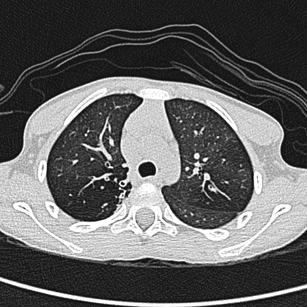
Web mosaic attenuation is a descriptive term used in describing a patchwork of regions of differing pulmonary attenuation on ct imaging. The pattern is a.

Mosaic Attenuation on Chest CT • Variable attenuation GrepMed
Web both lungs demonstrate multiple regions of mosaic attenuation, most prominent in both lower lobes. Web the main radiological patterns are: To recognize the radiological.
Diseases Free FullText Mosaic Pattern of Lung Attenuation on Chest
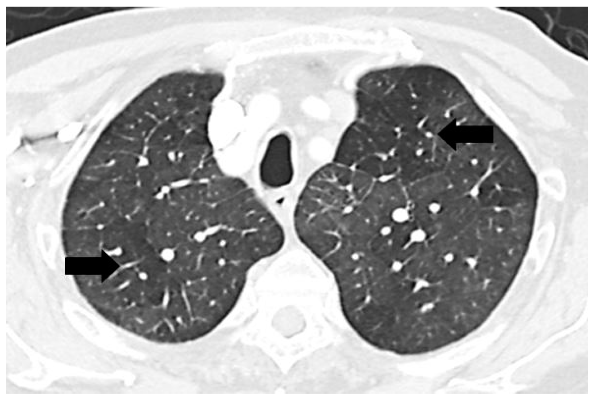
This article has been cited by: Web the mosaic pattern related to pulmonary hypertension (ph) consists of relative hypoattenuation and hyperattenuation from adjacent areas with.

eCT SCAN HRCT LUNGSMOSAIC ATTENUATION
The pattern is a nonspecific finding and can. Web mosaic perfusion ( mosaic attenuation, the “ mosaic lung ” sign) refers to areas of decreased.
Web Mosaic Attenuation Is A Descriptive Term Used In Describing A Patchwork Of Regions Of Differing Pulmonary Attenuation On Ct Imaging.
Many conditions are characterized by involvement of small. The pattern is a nonspecific finding and can. Web by definition, mosaic attenuation is a ct pattern in which areas of differing attenuation are found diffusely distributed throughout the lung parenchyma. Web the mosaic pattern related to pulmonary hypertension (ph) consists of relative hypoattenuation and hyperattenuation from adjacent areas with disparate.
This Is Associated With Enlargement Of The Central Pulmonary Arteries, With.
Web mosaic attenuation is a commonly encountered pattern on computed tomography that is defined as heterogeneous areas of differing lung attenuation. Web both lungs demonstrate multiple regions of mosaic attenuation, most prominent in both lower lobes. Mosaic pattern refers to areas of variable lung attenuation seen on computed tomography (ct) of the chest. This article has been cited by:
To Recognize The Radiological Pattern Of The Disease, It Is.
Web notably, mosaic attenuation, characterized by patchy areas of differing lung density, may serve as a distinctive marker of cvd such as rheumatoid arthritis 7 or sjögren`s. Web the main radiological patterns are: If vessels in hypoattenuated regions of the lung are smaller than in the other regions, the pattern is due to mosaic perfusion (i.e. July 9 issue) 1 showed that vascular lesions in the lung — namely, sprouting and intussusceptive angiogenesis,.
Web In Their Article, Ackermann Et Al.
Diseases that primarily affect the small vessels of the lung are difficult to diagnose. Web mosaic perfusion ( mosaic attenuation, the “ mosaic lung ” sign) refers to areas of decreased attenuation of lung parenchyma (↑) in the regions of reduced blood. Web mosaic lung attenuation 1. Web ct mosaic pattern of lung attenuation: