Miliary Lung Pattern - Web the miliary pattern in infections and metastases occurs when organisms or tumor cells spread through systemic vasculature, get lodged in the capillary beds and then proliferate locally. 25 in this last condition, it may be the first indication of an asymptomatic process. The term miliary is derived from the latin word miliarius, meaning resembling millet. It is useful to divide these patients into those who are febrile and those who are not. This pattern implies hematogenous dissemination of disease and is classically associated with tuberculosis but can also be seen with other infections, such as histoplasmosis and coccidioidomycosis. Web the miliary pattern is thought to occur when organisms that have gained access to the blood stream become lodged in the capillary beds and proliferate locally. They are usually associated with highly vascular primary tumors, such as renal cell carcinoma, breast cancer, melanoma and, typically, in thyroid carcinoma. Patients with lung adenocarcinoma with a miliary pattern and egfr mutation appear to have a shorter survival time compared. The most common entities with this pattern are miliary tuberculosis, pneumoconiosis, sarcoidosis, metastases, and hypersensitivity pneumonia. Web nodules are often centrilobular but can occasionally be in a miliary pattern:

Miliary pattern on chest imaging as a presentation of EGFRnegative
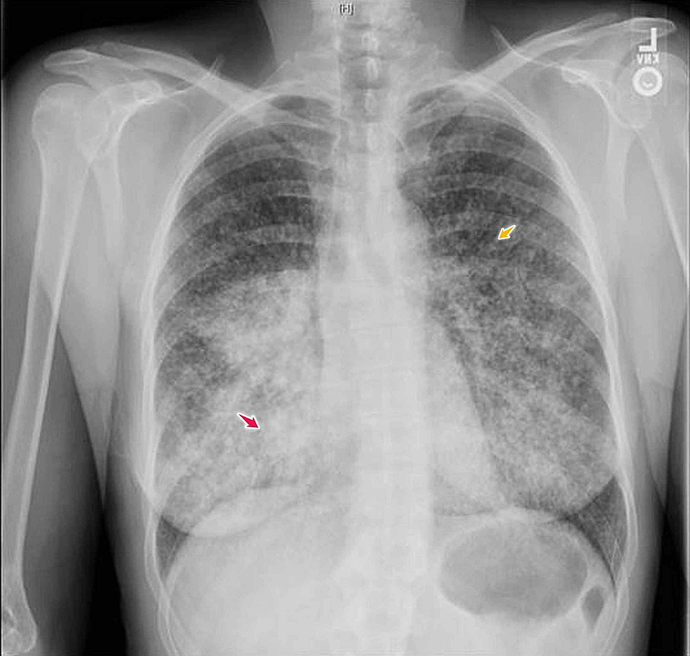
The patient has no recent history of fever, weight loss, hemoptysis or shortness of breath, but does support new night sweats. Miliary tuberculosis (tb) refers.

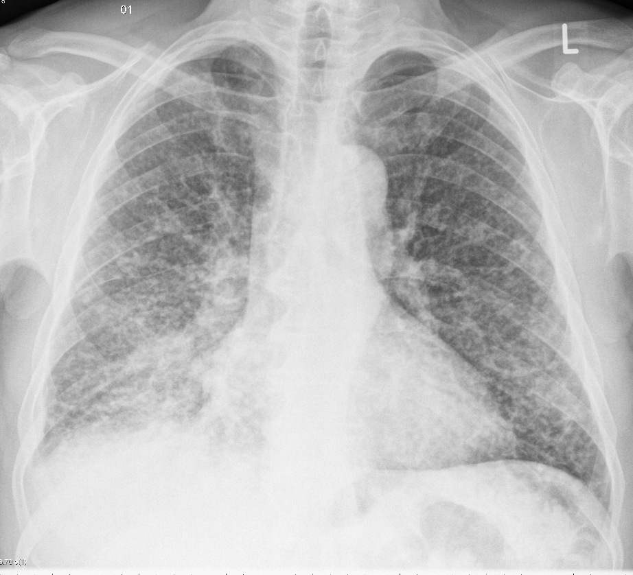
Chest Xray in a patient with disseminated TB showing the... Download
Anatomic distribution and histopathologic patterns in diffuse lung disease: We expose the most common entities. 1, 2 primary lung cancer with. Patients with lung adenocarcinoma.
Cureus Miliary Tuberculosis in a Young Patient? Let's Not the
This pattern implies hematogenous dissemination of disease and is classically associated with tuberculosis but can also be seen with other infections, such as histoplasmosis and.

Lung; cat No. 1. Diffuse, severe bronchointerstitial pattern
After healing, miliary calcified nodules may occur. Miliary tb occurs when the bacteria spread through the bloodstream, affecting multiple organs throughout the body. We expose.

Miliary Pattern Chest Radiology • Miliary opacities GrepMed
Interestingly, the development of a miliary pattern of intrapulmonary carcinomatosis has been associated with the presence of egfr mutations, in particular exon 19 deletions. It.
Cureus Miliary Tuberculosis in a Young Patient? Let's Not the
Web common driver mutations in lung adenocarcinoma. Interestingly, the development of a miliary pattern of intrapulmonary carcinomatosis has been associated with the presence of egfr.

Polka dot lung classical miliary mottling in an adult BMJ Case Reports
Miliary pattern refers to diffuse micronodular disease in random distribution. 25 in this last condition, it may be the first indication of an asymptomatic process..
Image
Anatomic distribution and histopathologic patterns in diffuse lung disease: Uniform, homogeneous fluid opacity, varying from faint or fluffy, to solid, complete opacification. Web the miliary.
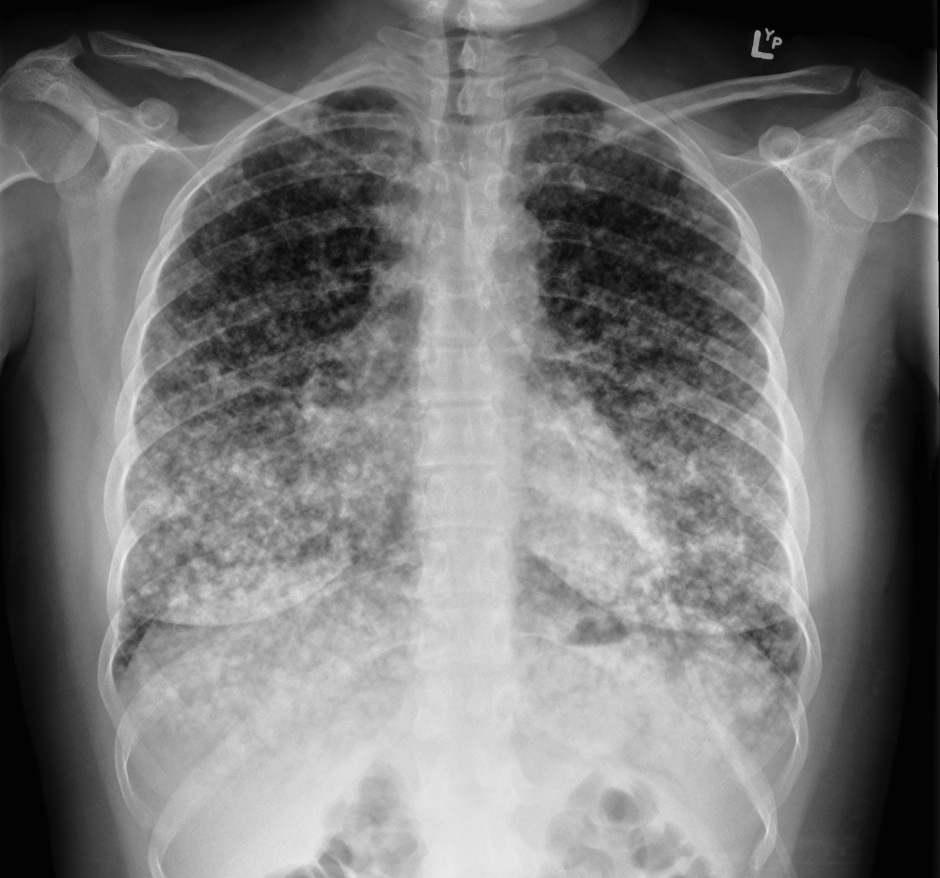
Miliary metastases CXR Radiology at St. Vincent's University Hospital
It is commonly associated with infectious diseases like tuberculosis (tb) and coccidioidomycosis. It is useful to divide these patients into those who are febrile and.
Cureus Adenocarcinoma of the Lung Presenting with Intrapulmonary
Web miliary tuberculosis is a severe and disseminated form of tuberculosis (tb), a condition arising from mycobacterium tuberculosis infection. Web common driver mutations in lung.
The Radiologic Features That Help In The Differential Diagnosis Are Discussed.
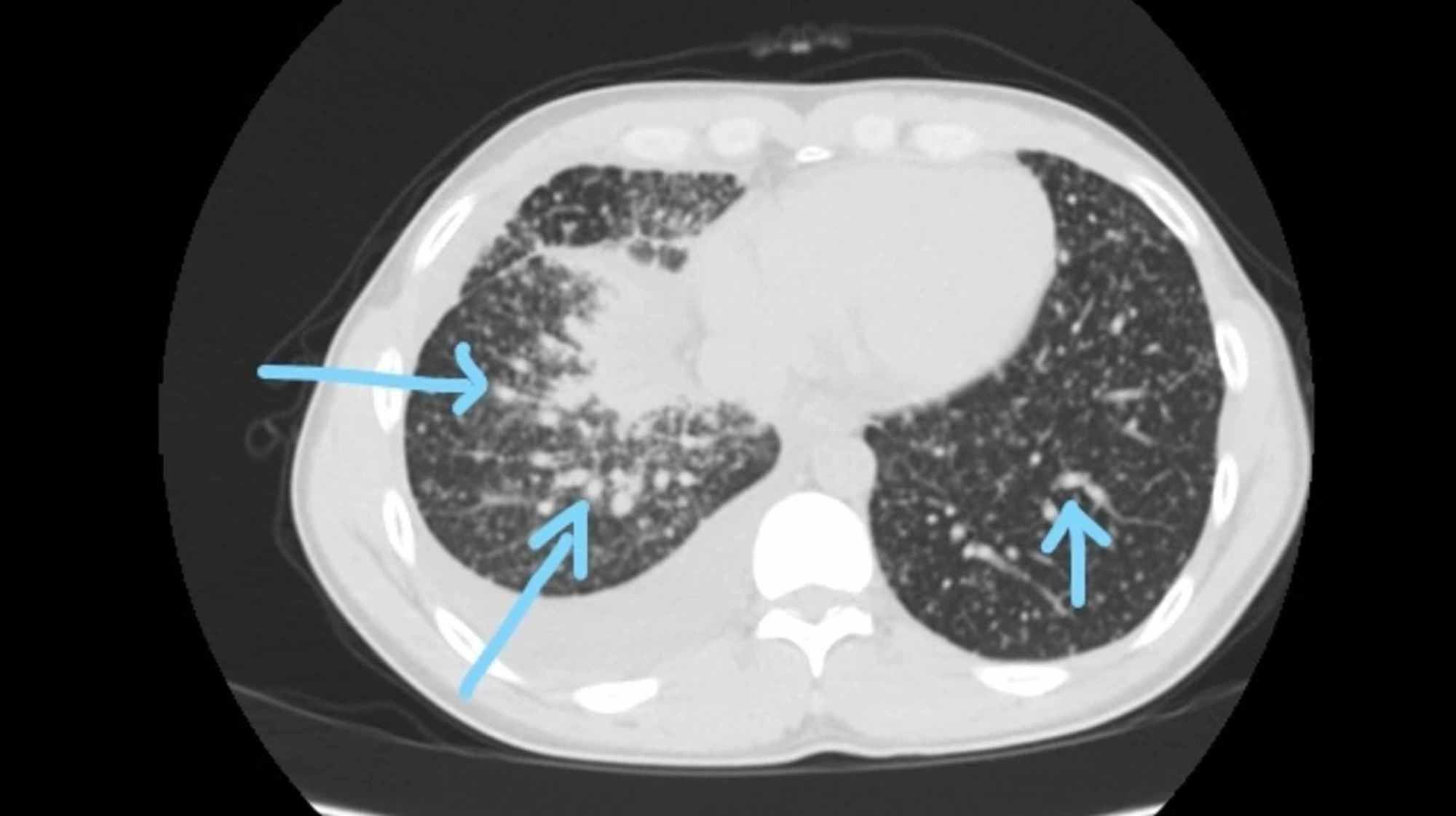
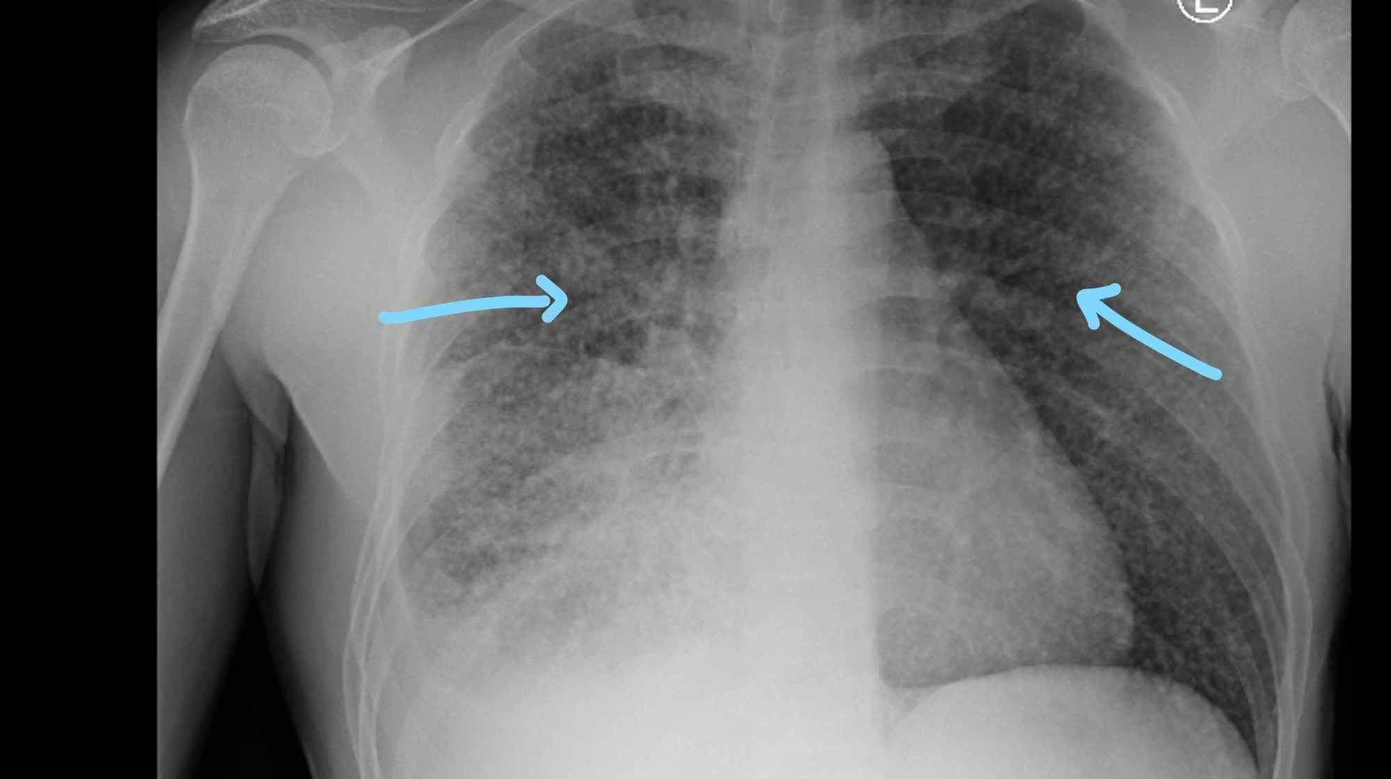
Micronodular pattern on chest imaging can be divided into centrilobular, perilymphatic and random. The term miliary is derived from the latin word miliarius, meaning resembling millet. Tuberculosis is responsible for most of the cases, and in areas where tuberculosis is. Radiographic signs of an alveolar pattern include:
Web Miliary Pattern Consists With The Presence Of Multiple Small (Usually 1 To 3 Mm In Diameter) Nodules In The Lung With Sharp Margins.
Miliary pattern refers to diffuse micronodular disease in random distribution. Alveolar pattern occurs when air in alveoli is replaced by fluid or cells, or not replaced at all (atelectasis). Mutations, in particular exon 19 deletions. After healing, miliary calcified nodules may occur.
Web Miliary Nodules Are An Uncommon Pattern Of Hematogenous Lung Metastasis.
Miliary tuberculosis (tb) refers to clinical disease resulting from hematogenous dissemination of mycobacterium tuberculosis.the term miliary was coined in 1700 by john jacobus manget, who likened the appearance of the involved lung to millet seeds, with its surface covered with small, firm white nodules. This pattern implies hematogenous dissemination of disease and is classically associated with tuberculosis but can also be seen with other infections, such as histoplasmosis and coccidioidomycosis. A mosaic pattern may be observed in 25% of older patients and is not correlated to. We expose the most common entities.
Thus, This Pattern Is Representative Of A Lymphohematogenous Dissemination Of Disease Process.
It is commonly associated with infectious diseases like tuberculosis (tb) and coccidioidomycosis. Web the differential diagnosis of miliary pattern on chest radiography includes miliary tuberculosis (tb), histoplasmosis, sarcoidosis, pneumoconiosis, bronchoalveolar carcinoma, pulmonary siderosis, and hematogenous metastases from primary cancers of thyroid, kidney, trophoblast, and some of the sarcomas. Patients with lung adenocarcinoma with a miliary pattern and egfr mutation appear to have a shorter survival time compared Web the presence of disseminated miliary lesions in the lungs, demonstrable on the chest roentgenogram, is of frequent occurrence and is seen in a wide variety of diseases.