Lung Patterns Dogs - Web these characteristic opacity changes are called lung patterns. Web the pattern approach to interpreting lung lesions simplifies your life. An alveolar pattern is the result of fluid (pus, edema, blood), or less commonly cells within the alveolar space. For reasons of simplicity we will not discuss mixed patterns. Web upper airway obstruction due to brachycephalic airway disease is a common cause of respiratory distress in brachycephalic dogs, such as english bulldogs. Web there are 4 pulmonary patterns described. Anthony fischetti, dvm, ms, dacvr, reviews the radiographic principles of lung patterns in dogs and cats. Identification of the lung pattern is helpful, as a list of differential diagnoses can be determined for that particular lung pattern. The lung appears diffusely more opaque or hazy. Examples of interstitial, alveolar, bronchial, and vascular lung patterns will be illustrated.

Photomicrographs of sections of the lung from the dog in Figure 1. AAn
Examples of interstitial, alveolar, bronchial, and vascular lung patterns will be illustrated. Radiographic abnormalities may help identify or suggest a primary cause of pulmonary hypertension;.

Dog lung lobes (from Dogs Monthly) Lung anatomy, Lunges, Dog anatomy
The clinical significance of these patterns will be discussed. Web see table 1 for differential diagnosis for common lung patterns in dogs and cats. Cardiogenic.

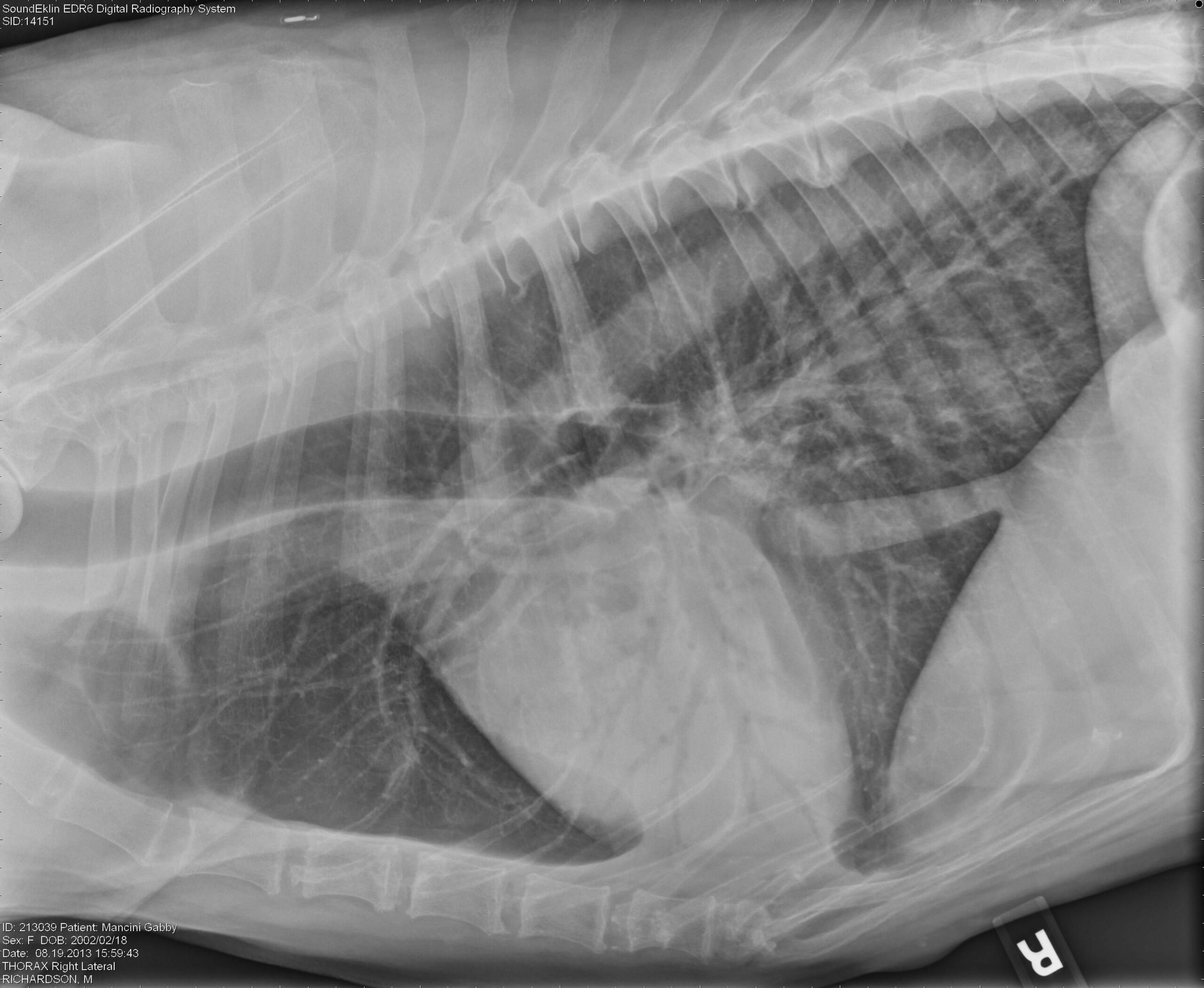
Thoracic radiographs of the canine patient. An interstitial pattern was
However, advanced imaging or additional diagnostic testing is necessary to confirm a diagnosis. For reasons of simplicity we will not discuss mixed patterns. Web there.

Radiographic Approach to the Coughing Pet MSPCAAngell
Radiographic abnormalities may help identify or suggest a primary cause of pulmonary hypertension; •application of lung patterns to common clinical. Lymphoma in dogs, primary pulmonary.

Common Pulmonary Diseases in Dogs Clinician's Brief
Anthony fischetti, dvm, ms, dacvr, reviews the radiographic principles of lung patterns in dogs and cats. Web canine and feline lungs have identical lobation with.
Interpreting thoracic radiograph lung patterns VETgirl Veterinary
Viral, bacterial or fungal) atelectasis (detected by the mediastinal shift when the alveoli are empty) bronchial. However, advanced imaging or additional diagnostic testing is necessary.

Thoracic radiography of a dog with pneumonic plague (case 2). Left
Web upper airway obstruction due to brachycephalic airway disease is a common cause of respiratory distress in brachycephalic dogs, such as english bulldogs. Web most.

Topographical distribution and radiographic pattern of lung lesions in
Structured and unstructured increased opacities. In cats, single circumscribed mass lesions are less common, whereas a diffuse lung pattern or. Examples of interstitial, alveolar, bronchial,.

Chronic & Persistent Coughing in a Dog Clinician's Brief
Web common lung patterns include: Structured and unstructured increased opacities. Identification of the lung pattern is helpful, as a list of differential diagnoses can be.

Topographical distribution and radiographic pattern of lung lesions in
Anthony fischetti, dvm, ms, dacvr, reviews the radiographic principles of lung patterns in dogs and cats. Web •review of lung patterns. Structured and unstructured increased.
Web The Pattern Approach To Interpreting Lung Lesions Simplifies Your Life.
Web •review of lung patterns. The contrast between the lung and vessels is diminished, and the vascular markings are poorly marginated. Emphasis will be placed on the diagnostic evaluation, paying special attention to interpretation of imaging findings and discrimination of these. Web most common in chronic lung diseases but also in neoplasia, toxic inhalation.
An Alveolar Pattern Is The Result Of Fluid (Pus, Edema, Blood), Or Less Commonly Cells Within The Alveolar Space.
Cardiogenic pulmonary edema is a common cause of respiratory distress in small breed dogs with chronic valvular disease (eg, mitral endocardiosis), such as cavalier king charles spaniels. Web upper airway obstruction due to brachycephalic airway disease is a common cause of respiratory distress in brachycephalic dogs, such as english bulldogs. •application of lung patterns to common clinical. Web dogs and cats with respiratory tract disorders can present to veterinarians for a variety of clinical signs including nasal discharge, sneeze, reverse sneeze, noisy breathing (snoring/stertor, stridor, wheeze), cough, alterations in respiratory rate or effort, and respiratory distress.
Web There Are 4 Pulmonary Patterns Described.
Structured and unstructured increased opacities. Web see table 1 for differential diagnosis for common lung patterns in dogs and cats. However, advanced imaging or additional diagnostic testing is necessary to confirm a diagnosis. Web these characteristic opacity changes are called lung patterns.
Web Canine And Feline Lungs Have Identical Lobation With Four Lobes Of The Right Lung (The Cranial, Middle, Caudal, And Accessory Lobes) And Two Lobes Of The Left Lung (The Cranial And Caudal Lobes).
A total collapse of the alveoli (atelectasis) leads to a. Web common lung patterns include: Examples of interstitial, alveolar, bronchial, and vascular lung patterns will be illustrated. Interstitial patterns indicate disease or disruption of the interstitium.