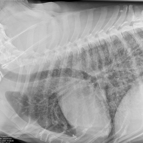
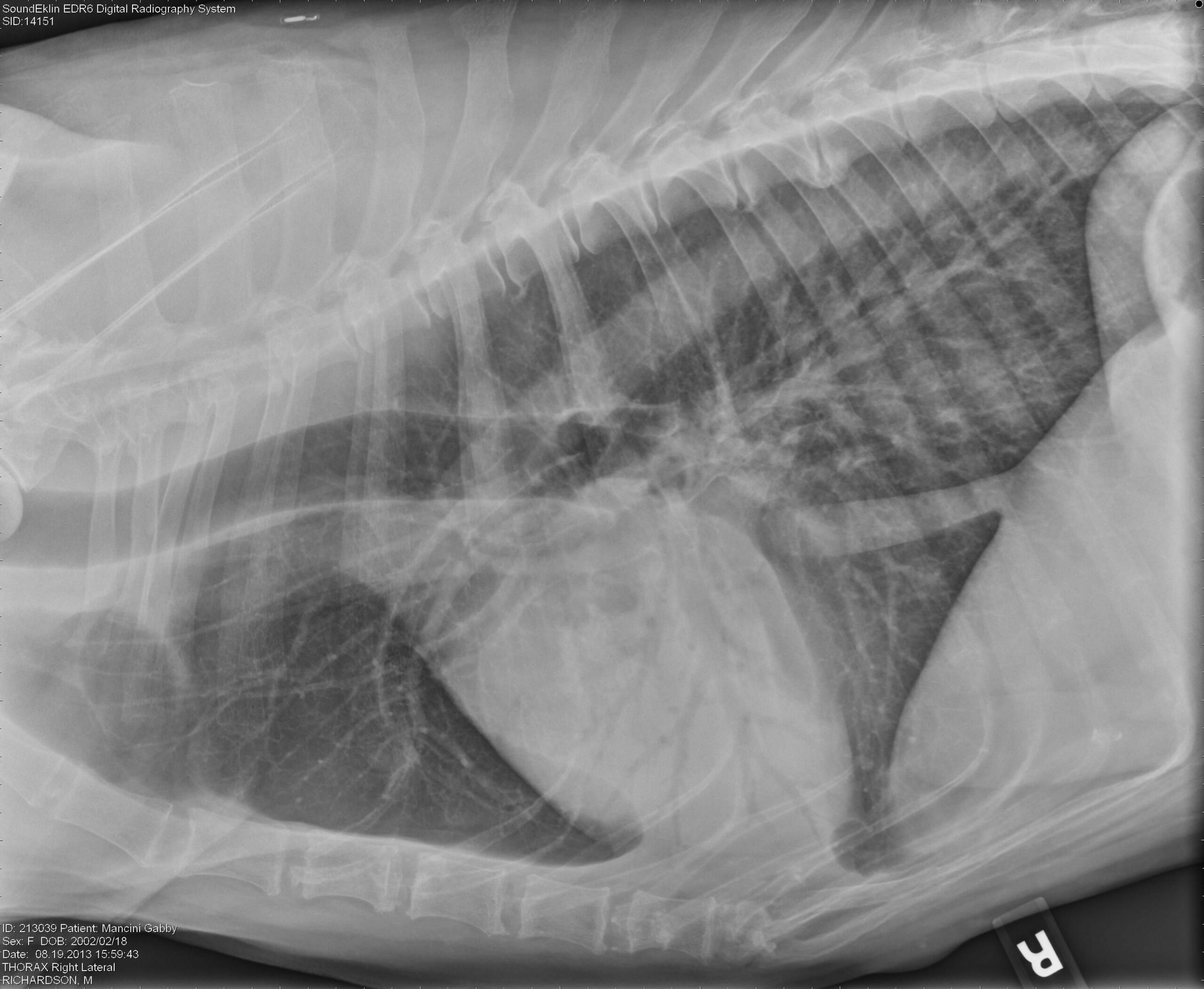
Lung Patterns Dog - Web learn how to differentiate pleural effusion and pulmonary edema on thoracic radiographs based on changes in opacity, fissure lines, and cardiac silhouette. Radiography remains the primary imaging modality for assessment of the thorax in first opinion practice. Web common lung patterns include: Web upper airway obstruction due to brachycephalic airway disease is a common cause of respiratory distress in brachycephalic dogs, such as english bulldogs. An unstructured interstitial pattern is present in the dorsocaudal lung fields structured interstitial (nodular) pattern. Characteristic findings include an increased opacity in the lungs that partially obscures blood vessel margins, which may be due to the presence of edema, pus, blood or other material in the lungs. The pattern approach to interpreting lung lesions simplifies your life. Web pulmonary disease is a common cause of respiratory signs in dogs. Interstitial patterns indicate disease or disruption of the interstitium. Web lateral thoracic radiograph of a dog with mitral insufficienty and interstital pulmonary edema.

Topographical distribution and radiographic pattern of lung lesions in
Web there are 4 pulmonary patterns described. An unstructured interstitial pattern is present in the dorsocaudal lung fields structured interstitial (nodular) pattern. Web respiratory distress.
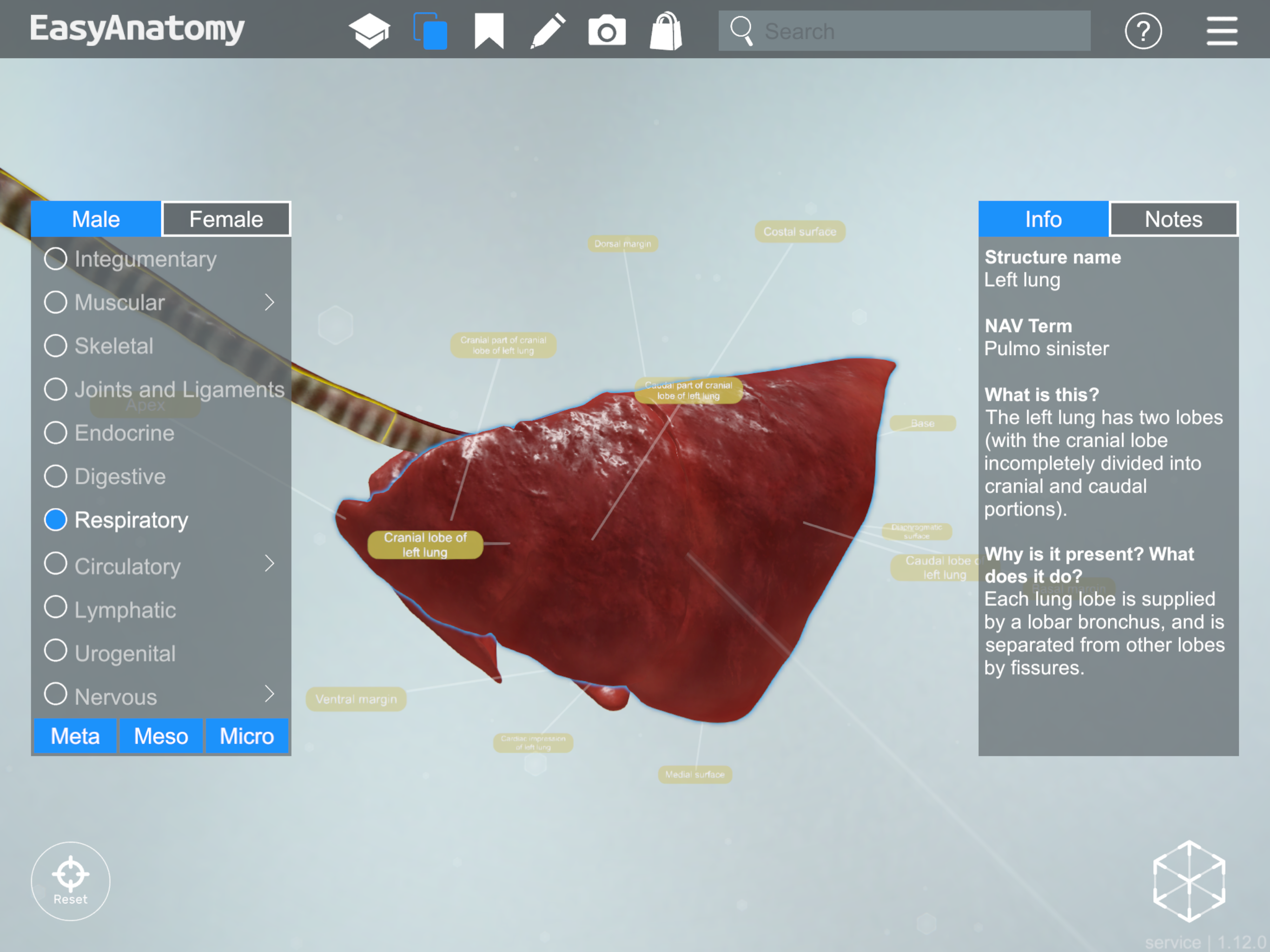
Anatomy of the Canine Respiratory System EasyAnatomy
The article describes the normal alveolar, bronchial, vascul… There is a wide variation in how the lung appears based on age and body condition in.
Common Pulmonary Diseases in Dogs Clinician's Brief
Web common lung patterns include: Characteristic findings include an increased opacity in the lungs that partially obscures blood vessel margins, which may be due to.

Photomicrographs of sections of the lung from the dog in Figure 1. AAn
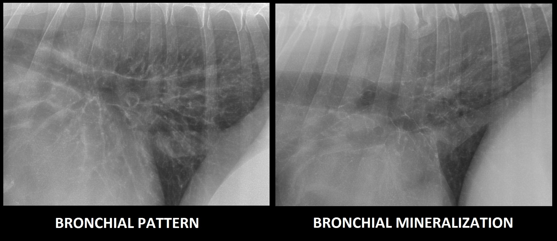
A thorough history and examination help localize the pulmonary parenchyma rather than other parts of the respiratory system. Web the bronchial pattern is most common.

Topographical distribution and radiographic pattern of lung lesions in
Their airways are very small, so look in the periphery for donuts and especially on the v/d or d/v projection where they show up better..
Interpreting thoracic radiograph lung patterns VETgirl Veterinary
Characteristic findings include an increased opacity in the lungs that partially obscures blood vessel margins, which may be due to the presence of edema, pus,.

Dog lung lobes (from Dogs Monthly) Lung anatomy, Lunges, Dog anatomy
Characteristic findings include an increased opacity in the lungs that partially obscures blood vessel margins, which may be due to the presence of edema, pus,.

Dog lung, illustration Stock Image F025/6691 Science Photo Library
Assigning a radiographic abnormality to a specific lung lobe is crucial to be able to narrow down the list of differential diagnoses. Characteristic findings include.
Radiographic Approach to the Coughing Pet • MSPCAAngell
In cats, single circumscribed mass lesions are less common, whereas a diffuse lung pattern or. Their airways are very small, so look in the periphery.

Radiographic Approach to the Coughing Pet • MSPCAAngell
Signalment, clinical onset and progression, geographic location, and additional organ involvement can help prioritize differential diagnoses. For reasons of simplicity we will not discuss mixed.
Learn How To Recognize And Interpret The Common Lung Patterns In Dogs Using Radiographic Imaging.
Radiography remains the primary imaging modality for assessment of the thorax in first opinion practice. There is a wide variation in how the lung appears based on age and body condition in normal animals. Clinically when faced with a mixed pattern, identify the most severe ( i.e. A thorough history and examination help localize the pulmonary parenchyma rather than other parts of the respiratory system.
In Cats, Single Circumscribed Mass Lesions Are Less Common, Whereas A Diffuse Lung Pattern Or.
The pattern approach to interpreting lung lesions simplifies your life. Web pulmonary disease is a common cause of respiratory signs in dogs. Web learn how to differentiate pleural effusion and pulmonary edema on thoracic radiographs based on changes in opacity, fissure lines, and cardiac silhouette. Web learn how to use thoracic radiographs to differentiate pulmonary and cardiac diseases in dogs and cats.
The Article Describes The Normal Alveolar, Bronchial, Vascul…
The altered opacity of the lung may be either increased (more opaque) or decreased (more lucent), but the majority of pulmonary diseases in dogs and cats produce an increased opacity. Web lateral thoracic radiograph of a dog with mitral insufficienty and interstital pulmonary edema. Because the act of respiration changes the thoracic appearance, inspiratory films should be attempted to distinguish artifacts from true lung pathology. Create an account for free.
Web Upper Airway Obstruction Due To Brachycephalic Airway Disease Is A Common Cause Of Respiratory Distress In Brachycephalic Dogs, Such As English Bulldogs.
Web companion animal vol. Normal variants causing increased lung opacity expiration: Signalment, clinical onset and progression, geographic location, and additional organ involvement can help prioritize differential diagnoses. A magnifying glass can come in handy as well.