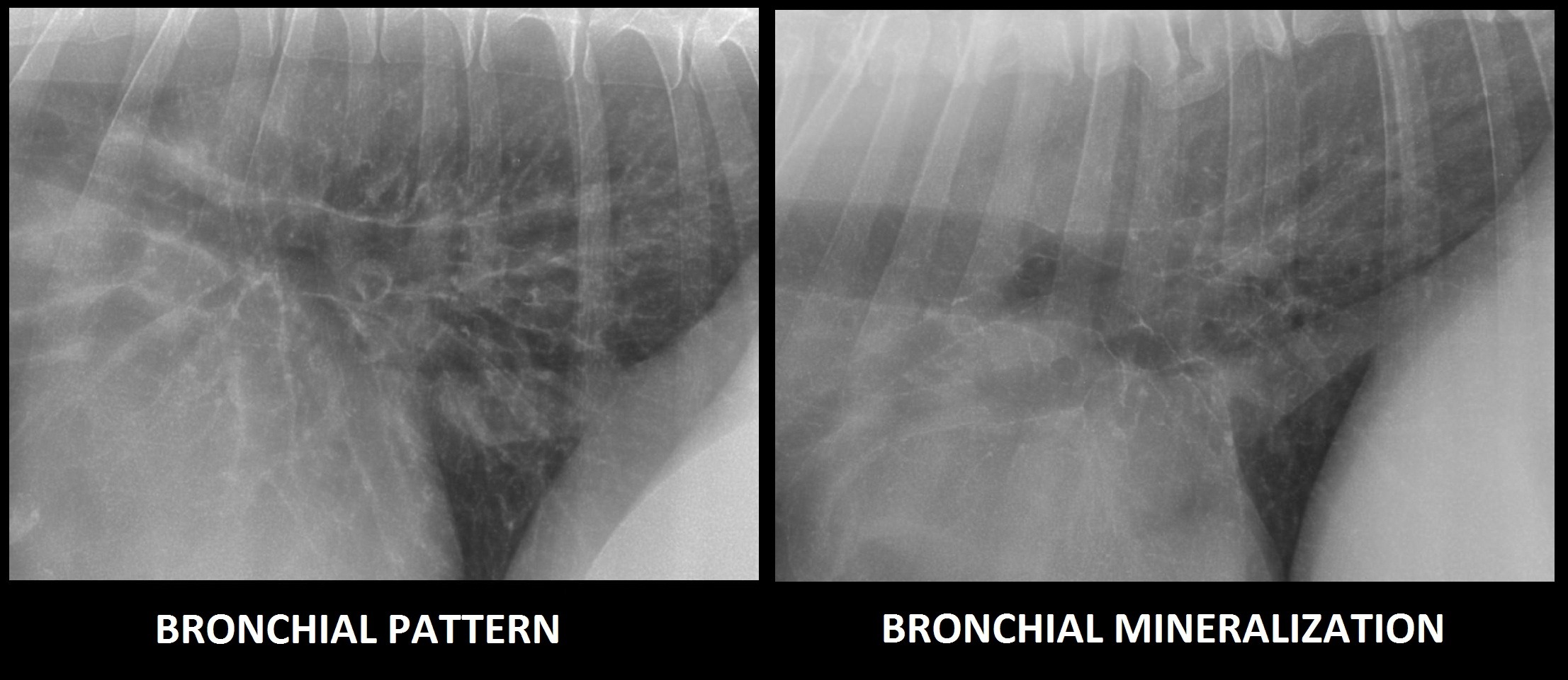
Lung Pattern Dog - The most reasonable conclusion is that the lesion is an alveolar pattern, but this is not based in either the presence of air bronchograms or a lobar sign. Web see table 1 for differential diagnosis for common lung patterns in dogs and cats. Because the act of respiration changes the thoracic appearance, inspiratory films should be attempted to distinguish artifacts from true lung pathology. Web common lung patterns include: Get expert advice90 day guarantee7,000+ positive reviews A bronchial pattern is diffuse thickening of the airway walls giving the appearance of thick lines and rings throughout the lungs. There is a wide variation in how the lung appears based on age and body condition in normal animals. 12mm+ questions answeredhelped over 8mm worldwide Interstitial patterns indicate disease or disruption of the interstitium. A bronchial and bronchointerstitial pattern are the most common radiographic lung patterns seen in canine eosinophilic bronchopneumopathy with these patterns most frequently topographically distributed to at least the caudodorsal lung field.
Interpreting thoracic radiograph lung patterns VETgirl Veterinary
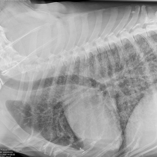
There are 4 pulmonary patterns described. In cats, single circumscribed mass lesions are less common, whereas a diffuse lung pattern or. Web common lung patterns.

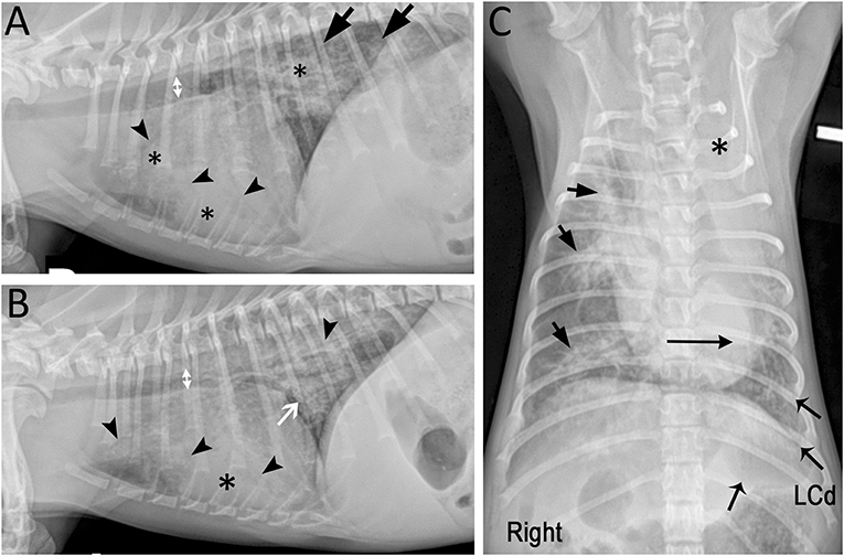
Topographical distribution and radiographic pattern of lung lesions in
There are 4 pulmonary patterns described. — cranioventral distributions tend to be associated with pneumonias. Matthew winter, dacvr will review the radiographic features of lung.
Radiographic Approach to the Coughing Pet • MSPCAAngell
Web common lung patterns include: The pattern approach to interpreting lung lesions simplifies your life. A bronchial and bronchointerstitial pattern are the most common radiographic.

Dog lung, illustration Stock Image F025/6691 Science Photo Library
A bronchial pattern is diffuse thickening of the airway walls giving the appearance of thick lines and rings throughout the lungs. Normal variants causing increased.

Dog lung lobes (from Dogs Monthly) Lung anatomy, Lunges, Dog anatomy
Download appcall or chat 24/7america's #1 pet pharmacywe contact your vet The altered opacity of the lung may be either increased (more opaque) or decreased.

Photomicrographs of sections of the lung from the dog in Figure 1. AAn
Matthew winter, dacvr will review the radiographic features of lung patterns in dogs and cats as well as the keys to interpreting the meaning of.

Veterinary Key Points Canine Lung Lobectomy Video
There is a wide variation in how the lung appears based on age and body condition in normal animals. Download appcall or chat 24/7america's #1.
Frontiers Presumptive Development of Fibrotic Lung Disease From
Web six regions from the vd views, and two from the lm views, were evaluated by three of the four study authors (all radiologists) and.

Topographical distribution and radiographic pattern of lung lesions in
Normal (p1), alveolar pattern (p2), bronchial pattern. Examples of this will be shown. Interstitial patterns indicate disease or disruption of the interstitium. Furthermore, within the.
Common Pulmonary Diseases in Dogs Clinician's Brief
Download appcall or chat 24/7america's #1 pet pharmacywe contact your vet Web the defining sign that helps us determine why the dog/cat can't breath is.
Web Primary Lung Tumors In Dogs May Occur As Single Or Multiple Circumscribed Mass Lesions, As A Diffuse Lung Pattern, Or As A Lobar Consolidation.
12mm+ questions answeredhelped over 8mm worldwide Download appcall or chat 24/7america's #1 pet pharmacywe contact your vet For reasons of simplicity we will not discuss mixed patterns. Trauma (such as being hit by a car) may.
An Unstructured Interstitial Pattern Is Present In The Dorsocaudal Lung Fields Structured Interstitial (Nodular) Pattern.
Get expert advice90 day guarantee7,000+ positive reviews The purpose of this article is to describe common principles of the radiological survey of the lungs, which is crucial for the making of correct and timely diagnosis in the clinical settings. Ventrodorsal radiograph of a normal dog; Furthermore, within the caudodorsal lung field, a bronchointerstitial pattern predominates.
Normal Variants Causing Increased Lung Opacity Expiration:
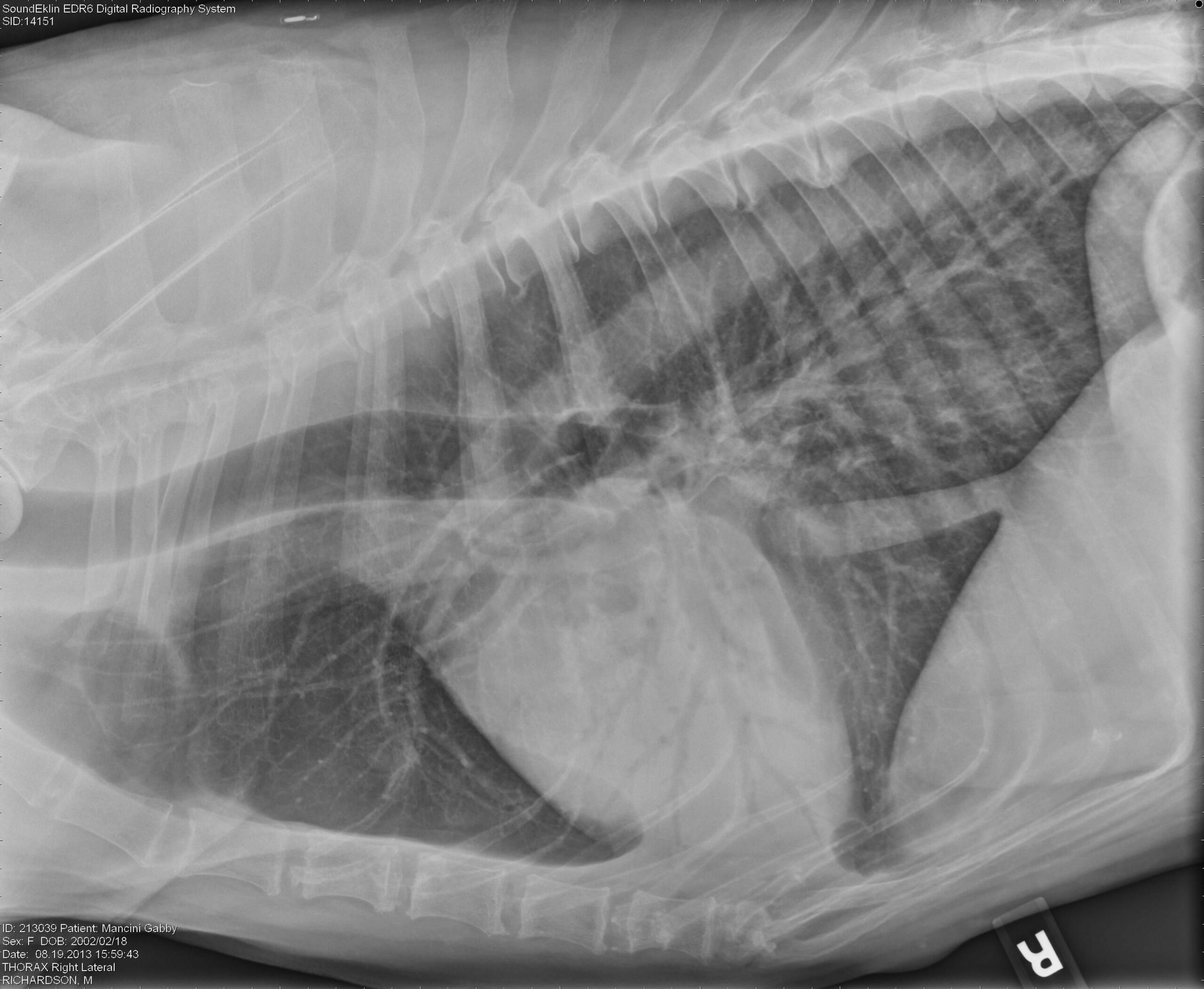
Interstitial patterns indicate disease or disruption of the interstitium. The pleural space exists between each lung lobe at the interlobar fissure as. Because the act of respiration changes the thoracic appearance, inspiratory films should be attempted to distinguish artifacts from true lung pathology. Web lateral thoracic radiograph of a dog with mitral insufficienty and interstital pulmonary edema.
In Cats, Single Circumscribed Mass Lesions Are Less Common, Whereas A Diffuse Lung Pattern Or.
Web typical differentials for interstitial and alveolar patterns in dogs include: A bronchial pattern is important to recognize, because, while it may be a normal variant in an aged. Vet talks is a project by the ivsa standing committee on. Create an account for free.