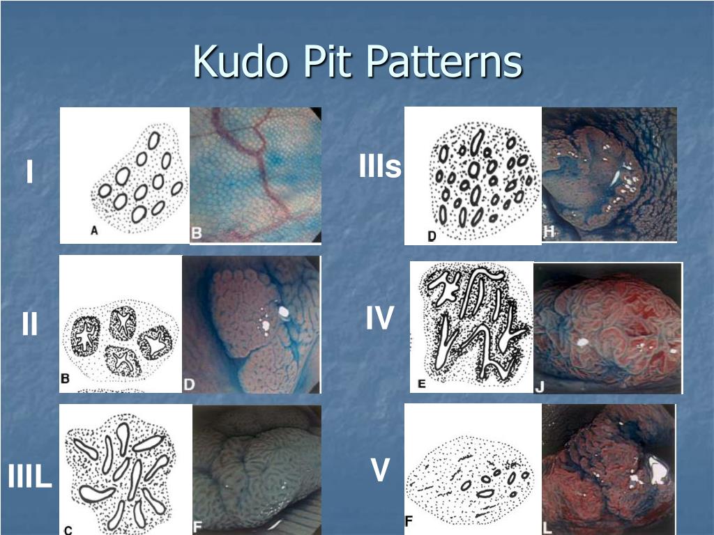
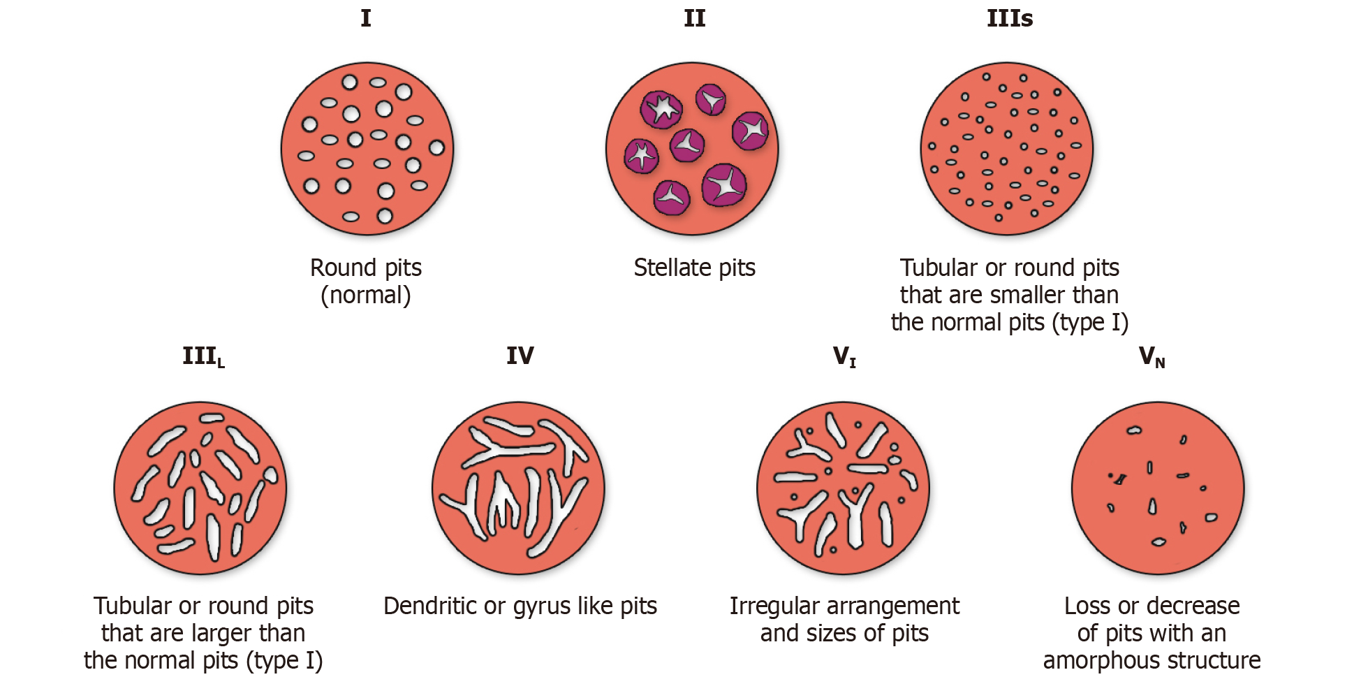
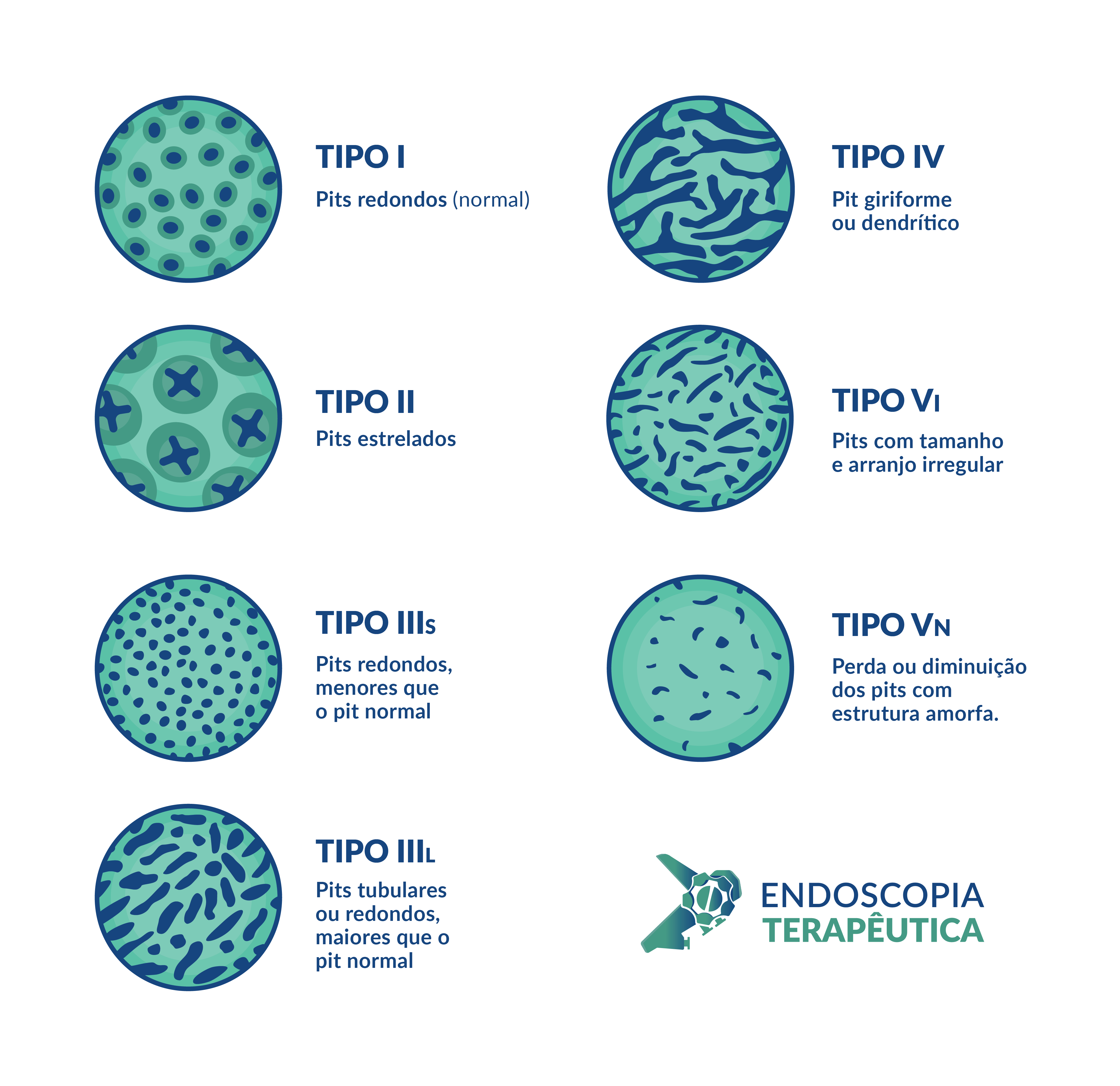
Kudo Pit Pattern - Here’s the kudo classification of polyps and their correlation with pathology: The kudo classification classifies pit pattern as round / normal (type i), asteroid (type ii), tubular or round pit smaller than normal pit (type iiis), tubular or round pit larger than normal pit (type iiil), gyrus/dendritic (iv), Publication bias is significant in the current available literature. This scheme broadly categorizes pit patterns into 7 types based on the pit appearance and structure (figure 3 ). Kudo and colleagues first highlighted the feasibility of examining and classifying pit patterns to distinguish type of polyps by using magnifying endoscopy. Web evaluating 2693 lesions, kudos pit pattern v was the strongest factor associated with overt submucosal invasive cancer (odds ratio [or], 1.42; Lesion stratification, architecture recognition and estimation of depth of invasion are crucial for patient selection. To analyze the current available evidence of kudo's pit pattern classification for diagnosing colorectal neoplasms. Adapted and reprinted from tanaka et al. Web kudo's pit pattern classification is an accurate diagnostic method for the differentiation of neoplastic colorectal lesions and publication bias is significant in the current available literature.

Figure 1 from Quantitative analysis and development of a computeraided
Lesion stratification, architecture recognition and estimation of depth of invasion are crucial for patient selection. Kudo and colleagues first highlighted the feasibility of examining and.

Classificação de Kudo Pit pattern Endoscopia Terapêutica
A search was performed on pubmed/embase to identify studies reporting the outcomes of the pit pattern classification in colorectal polyps. Web the kudo pit pattern.

Kudo's pit pattern classification was composed seven type pit pattern
Web the kudo pit pattern (figure 5), with the use of dye chromoendoscopy, has been found to be highly accurate for predicting superficial (type vi).

Classification of pit patterns of colorectal lesions. Download
[115], with permission from elsevier. Type ii includes stellate or papillary pits, which indicate hyperplasia. Web the kudo pit pattern (figure 5), with the use.
PPT EQUIP Training session 2 PowerPoint Presentation, free download
Web for microvascular pattern evaluation, the nbi international colorectal endoscopic (nice) classification and the japanese nbi expert team (jnet) classification are the most widely disseminated,.

Pit pattern classification of colorectal neoplasia (Adapted from Tanaka
Web a value of 4 or higher in the kudo classification turned out to be the best parameter to differentiate malignant lesions from benign ones.
Endoscopic management of colorectal polyps From benign to malignant polyps
Web for microvascular pattern evaluation, the nbi international colorectal endoscopic (nice) classification and the japanese nbi expert team (jnet) classification are the most widely disseminated,.
Classificação de Kudo Pit pattern • Endoscopia Terapeutica
A search was performed on pubmed/embase to identify studies reporting the outcomes of the pit pattern classification in colorectal polyps. Web kudo classification of pit.

Kudo classification of pit patterns with photographic correlation of
Kudo and colleagues first highlighted the feasibility of examining and classifying pit patterns to distinguish type of polyps by using magnifying endoscopy. The kudo classification.

Kudo classification of pit patterns with photographic correlation of
Web the kudo pit pattern classification system uses magnification colonoscopy with dye spray (chromoendoscopy) to categorize polyps based on the appearance of their pits, which.
This Scheme Broadly Categorizes Pit Patterns Into 7 Types Based On The Pit Appearance And Structure (Figure 3 ).
Web kudo's pit pattern classification is an accurate diagnostic method for the differentiation of neoplastic colorectal lesions. Web the kudo classification classifies pit pattern as round / normal (type i), asteroid (type ii), tubular or round pit smaller than normal pit (type iiis), tubular or round pit larger than normal pit (type iiil), gyrus/dendritic (iv), irregular arrangement (vi), and loss of decrease of pits with amorphous structure (vn). Web kudo's pit pattern classification is an accurate diagnostic method for the differentiation of neoplastic colorectal lesions and publication bias is significant in the current available literature. [115], with permission from elsevier.
Here’s The Kudo Classification Of Polyps And Their Correlation With Pathology:
15 the different pit patterns include round/normal (type i), asteroid (type ii), tubular or round pit that is smaller than the normal (type iiis. 6 type i includes round pits that are observed in normal mucosa. Adapted and reprinted from tanaka et al. Web the kudo pit pattern classification system uses magnification colonoscopy with dye spray (chromoendoscopy) to categorize polyps based on the appearance of their pits, which are crypt openings.
Publication Bias Is Significant In The Current Available Literature.
Publication bias is significant in the current available literature. Accurate assessment of polyp size is one of the most important factors in determining the risk of cancer in a polyp. 12 however, assessing the kudo pit pattern has limited use owing to dye availability, additional procedural time for their preparation and application and. Web evaluation of the pit pattern of the polyps.
Even Polyps Of <10 Mm Have A Malignant Potential And This Can Be Largely Determined By A Careful Endoscopic Evaluation.
Kudo and colleagues first highlighted the feasibility of examining and classifying pit patterns to distinguish type of polyps by using magnifying endoscopy. Web kudo classification of pit patterns with photographic correlation of lesions. Web the kudo pit pattern (figure 5), with the use of dye chromoendoscopy, has been found to be highly accurate for predicting superficial (type vi) and deep submucosal invasion (type vn). Lesion stratification, architecture recognition and estimation of depth of invasion are crucial for patient selection.