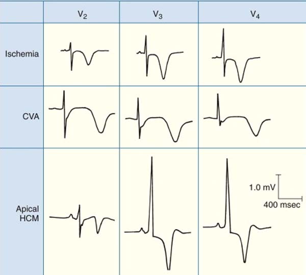
Juvenile T Wave Pattern - Should be upright in leads v5 and v6 in a normal ecg; Persistent juvenile t wave pattern. Uptodate, electronic clinical resource tool for physicians and patients that provides information on adult primary care and internal medicine, allergy and immunology, cardiovascular medicine. Web juvenile t wave pattern refers to the t wave inversions commonly seen in the anterior leads. It is characterized by twi in the right precordium, and has been understood to represent an arrested stage of the normal electrocardiographic evolution. Type i had negative t waves in lead v 1 only, type ii in lead v 1 and v 2 and type iii in v 1 to lead v 3 or v 4. Hyperkalaemia, lvh (volume overload), cerebral vascular accident. It is characterized by twi in the right precordium, and has been understood to represent an arrested stage of the normal electrocardiographic evolution. The three dynamical phases are: Type a = biphasic t waves with.
Dr. Smith's ECG Blog Persistent Juvenile Twave Pattern
For example, the whales would systematically modulate certain aspects of their codas based on the. It is characterized by twi in the right precordium, and.
Dr. Smith's ECG Blog Persistent Juvenile Twave Pattern
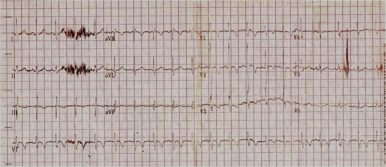
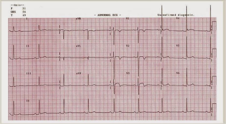
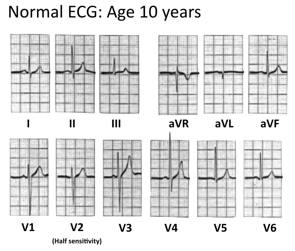
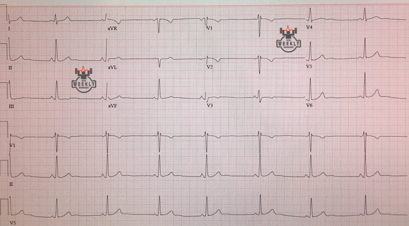
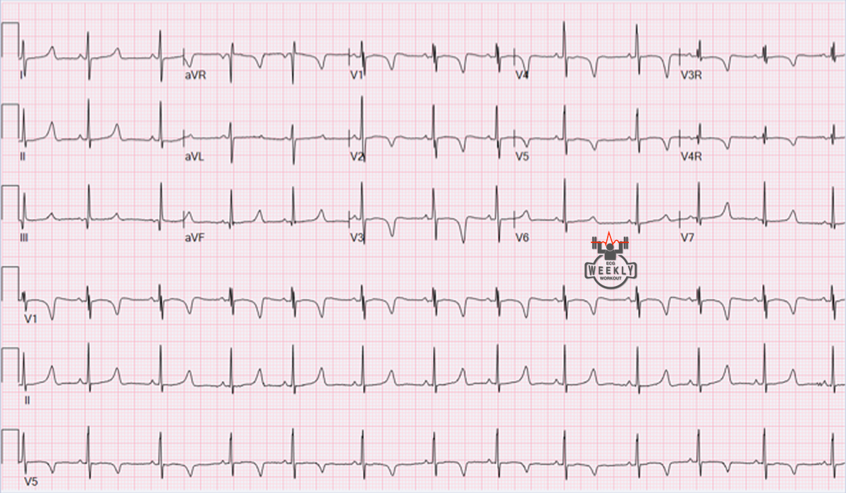
Web the t waves in the right precordial leads (v1 through v3) are asymmetrically inverted in childhood and become upright (except for v1 and occasionally.
Persistent Juvenile T Wave Pattern ECG Stampede
Hyperkalaemia, lvh (volume overload), cerebral vascular accident. In predominantly black women under the age of 40, this juvenile t wave pattern may persist in the.
Paediatric ECG Interpretation • LITFL • ECG Library Basics
It is characterized by twi in the right precordium, and has been understood to represent an arrested stage of the normal electrocardiographic evolution. Type i.
ECG T wave changes and interpretation Medicine Hack
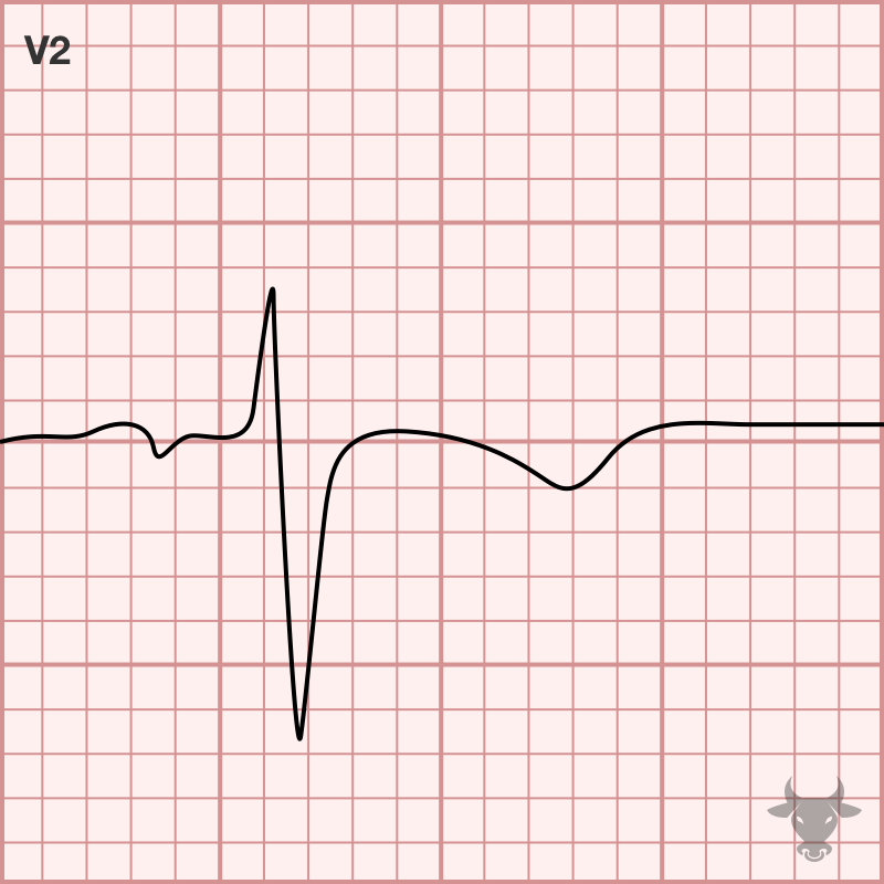
Should be upright in leads v5 and v6 in a normal ecg; These inversions show a slow descent, with a relatively brisk upstroke, and there.
Persistent juvenile Twave pattern ECG Weekly
Persistent juvenile t wave patterns found in mass examination and in clinical cases were studied for the purpose of clarifying their significance. Anterior twi, however,.

“Persistent Juvenile” TWave Pattern May Not Be Persistent Case Series
It is characterized by twi in the right precordium, and has been understood to represent an arrested stage of the normal electrocardiographic evolution. Type i.
Juvenile Twave pattern ECG Weekly
It is characterized by twi in the right precordium, and has been understood to represent an arrested stage of the normal electrocardiographic evolution. Should be.

Dr. Smith's ECG Blog Persistent Juvenile Twave Pattern
Web the researchers identified something of a “sperm whale phonetic alphabet,” where various elements that researchers call “rhythm,” “tempo,” “rubato,” and “ornamentation” interplay to form.

The Twave physiology, variants and ECG features EKG & ECHO
Rsr’ pattern (partial rbbb morphology) in v1. Type i had negative t waves in lead v 1 only, type ii in lead v 1 and.
It Is Characterized By Twi In The Right Precordium, And Has Been Understood To Represent An Arrested Stage Of The Normal Electrocardiographic Evolution.
(emily smith/cnn) a stunning aurora, caused by a severe geomagnetic storm, is painting the sky shades of pink, purple and green as it spreads into. The changes may be seen in all or most of the leads (diffuse changes), or they may be present contiguous leads, such as the inferior, lateral, or anterior leads. Should be upright in leads v5 and v6 in a normal ecg; Case series and literature review,.
Web Juvenile T Wave Pattern Refers To The T Wave Inversions Commonly Seen In The Anterior Leads.
In predominantly black women under the age of 40, this juvenile t wave pattern may persist in the absence of cardiac abnormalities. Web the persistent juvenile t wave pattern consists of asymmetric t wave inversion seen in v1 through to v3 or v4. Persistent juvenile t wave patterns found in mass examination and in clinical cases were studied for the purpose of clarifying their significance. Rsr’ pattern (partial rbbb morphology) in v1.
Web The T Waves In The Right Precordial Leads (V1 Through V3) Are Asymmetrically Inverted In Childhood And Become Upright (Except For V1 And Occasionally V2) By Adulthood.
The types of abnormalities are varied and include subtle straightening of the st. It is named because it resembles the normal child's ecg [62, 63]. In the first week of life, the t waves are all upright. Type a = biphasic t waves with.
It Is Characterized By Twi In The Right Precordium, And Has Been Understood To Represent An Arrested Stage Of The Normal Electrocardiographic Evolution.
It is characterized by twi in the right precordium, and has been understood to represent an arrested stage of the normal electrocardiographic evolution. These inversions show a slow descent, with a relatively brisk upstroke, and there is no associated st segment deviation (fig. The three dynamical phases are: Uptodate, electronic clinical resource tool for physicians and patients that provides information on adult primary care and internal medicine, allergy and immunology, cardiovascular medicine.