In An Ecg Pattern The P Wave Is Caused By - This is associated with a delta wave. The pr interval begins at the start of the p. In an ecg pattern, the p wave is caused. Dilation of the right atrium and right ventricle with consequent shift in the position of the heart. If the left atrium encounters increased resistance (e.g. Web p waves are the key to determining whether a patient is in sinus rhythm or not. Web in an ecg pattern the p wave is caused by. Arterial systolic pressure is most closely associated with. Web it is caused by an impulse discharged from an ectopic focus which may be located anywhere in the atria. Ecg changes in pe are related to:

Pwave, PR interval, PR segment physiology, criteria & ECG findings
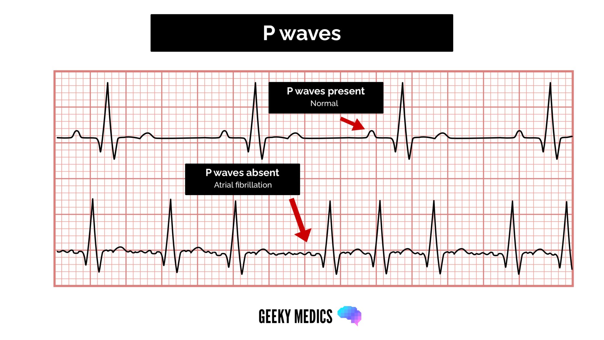
In healthy individuals, there should be a p wave preceding each qrs complex. This article is part of the comprehensive chapter: Sinus rhythm (which is.

Pwave, PR interval, PR segment physiology, criteria & ECG findings
Web the p wave and pr segment is an integral part of an electrocardiogram (ecg). Web p waves are the key to determining whether a.

The Electrocardiogram explained What is an ECG?
Web a p wave on an electrocardiogram represents a phase of electrical activity that causes the atria of the heart to contract. Web in an.

ECG interpretation Characteristics of the normal ECG (Pwave, QRS
Arterial systolic pressure is most closely associated with. Dilation of the right atrium and right ventricle with consequent shift in the position of the heart..

ECG interpretation Characteristics of the normal ECG (Pwave, QRS
Web the factors that determine p‐wave appearance include (1) the origin of the sinus rhythm that defines right atrial depolarization vector, (2) localization of left.
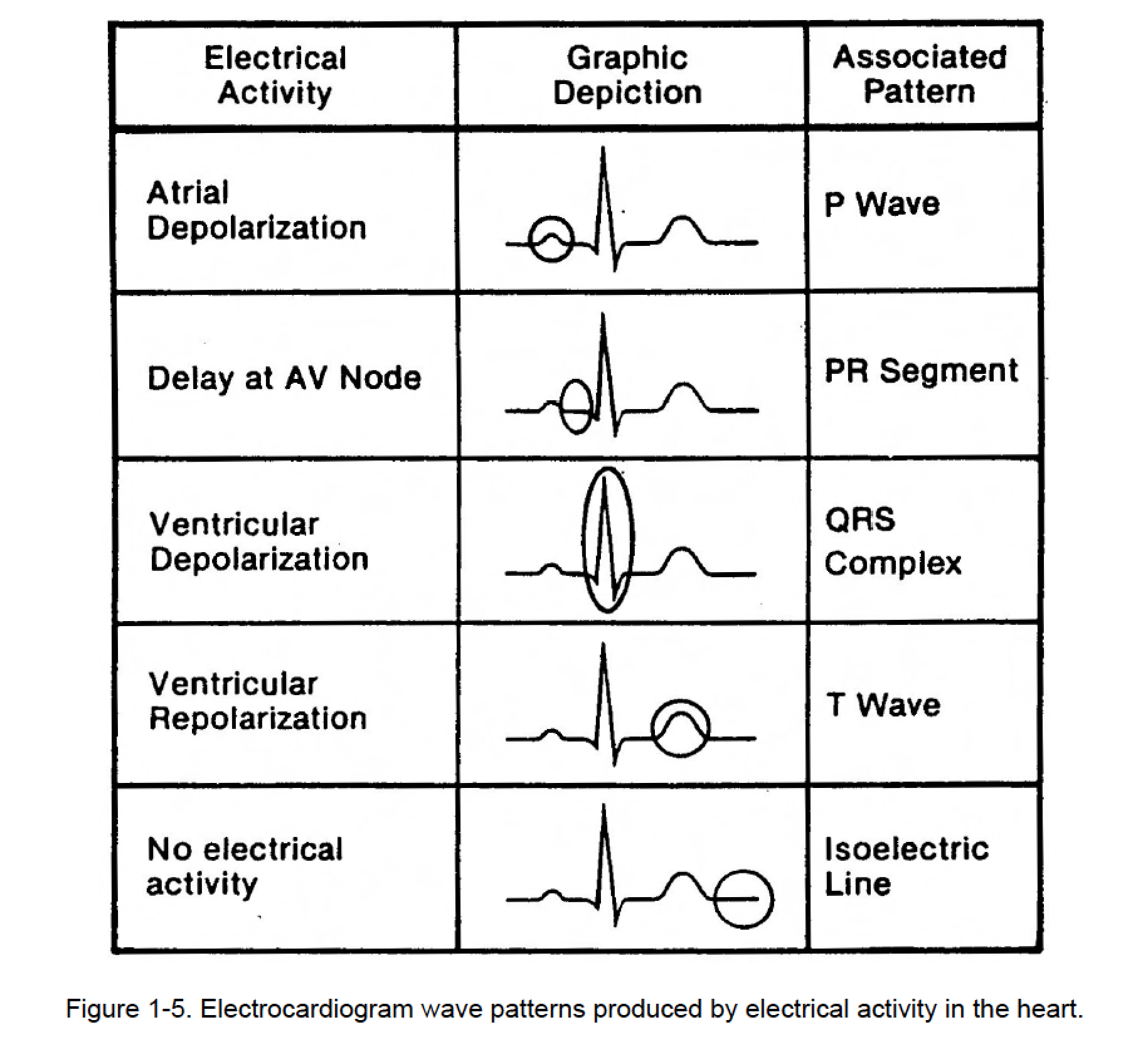
Figure 15. Cardiac Rhythm Interpretation
This article is part of the comprehensive chapter: Web study with quizlet and memorize flashcards containing terms like which of the following is not true.

Interpretation of the P wave YouTube
Web a p wave on an electrocardiogram represents a phase of electrical activity that causes the atria of the heart to contract. Web in an.
Electrocardiograms (EKGs/ECGs) Evaluating P Waves Stepwards
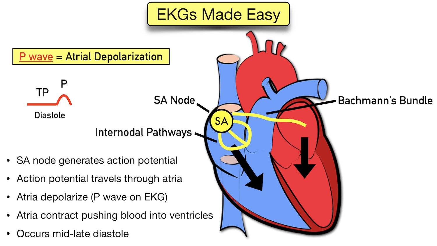
Web the p wave on the ecg represents atrial depolarization, which results in atrial contraction, or atrial systole. Web hyperkalaemia creates the illusion that the.
P wave abnormalities ecg ppt
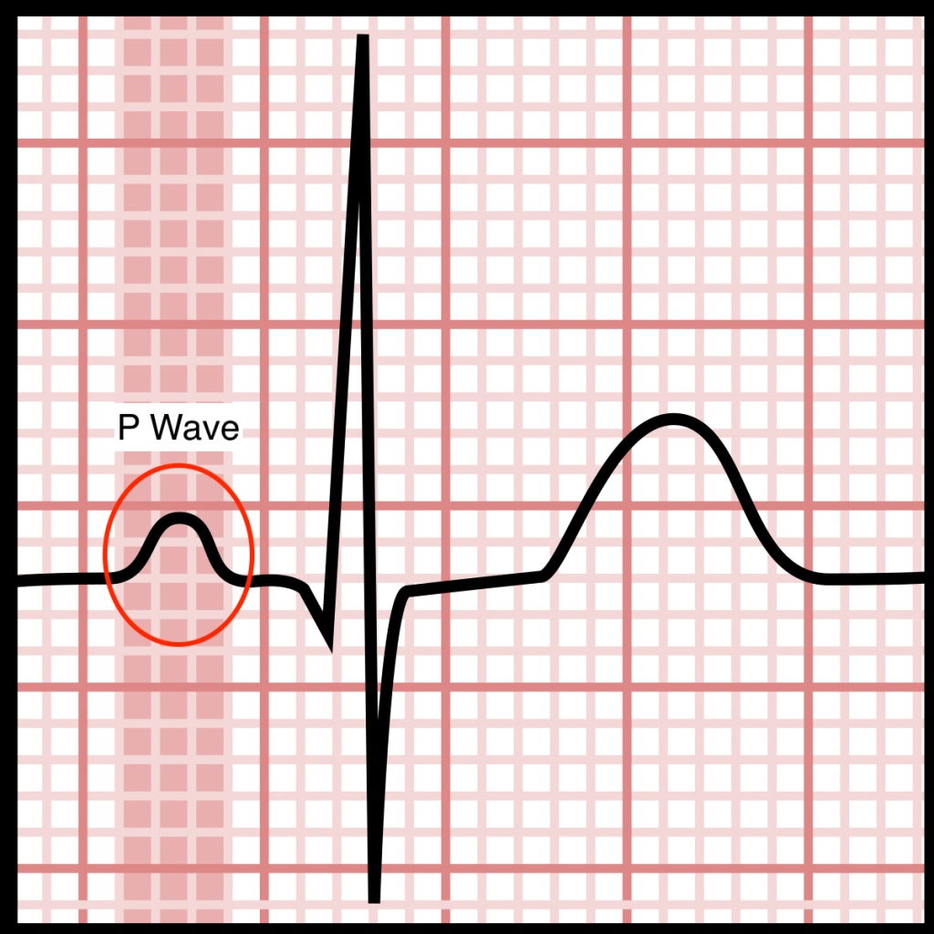
The p wave is a summation wave generated. This webpage explains the p wave, which represents atrial depolarization on the ecg, and how to. It.
ECG Waveform Explained EKG Labeled Diagrams and Components — EZmed
Web hyperkalaemia creates the illusion that the t wave is “pulled upwards”, creating tall “tented” t waves, and stretching the remainder of the ecg to.
It Is Typically A Small.
In healthy individuals, there should be a p wave preceding each qrs complex. Sinus rhythm (which is the normal rhythm) has the following characteristics: It represents the electrical depolarization of the atria of the heart. Arterial systolic pressure is most closely associated with.
Web In The Ecg Pattern, The Pq Interval Indicates How Long It Takes For The Cardiac Impulse To Travel From The.
Depolarization of atrial muscle fibers. Web hyperkalaemia creates the illusion that the t wave is “pulled upwards”, creating tall “tented” t waves, and stretching the remainder of the ecg to cause p wave flattening, pr. Web atrial flutter with a 3:1 block. If the left atrium encounters increased resistance (e.g.
Web P Waves Are The Key To Determining Whether A Patient Is In Sinus Rhythm Or Not.
Sa node through the av node. Web study with quizlet and memorize flashcards containing terms like which of the following is not true about the heart?, when the ventricular walls contract, which of the following. Web a typical ecg tracing of the cardiac cycle (heartbeat) consists of a p wave (atrial depolarization ), a qrs complex (ventricular depolarization), and a t wave (ventricular. As atrial depolarization initiates by the sa node located in the right atrium, the right atrium gets depolarized first, followed by.
The Pr Interval Begins At The Start Of The P.
Web it is caused by an impulse discharged from an ectopic focus which may be located anywhere in the atria. Ecg changes in pe are related to: It represents atrial depolarization on the ecg. Web a p wave on an electrocardiogram represents a phase of electrical activity that causes the atria of the heart to contract.