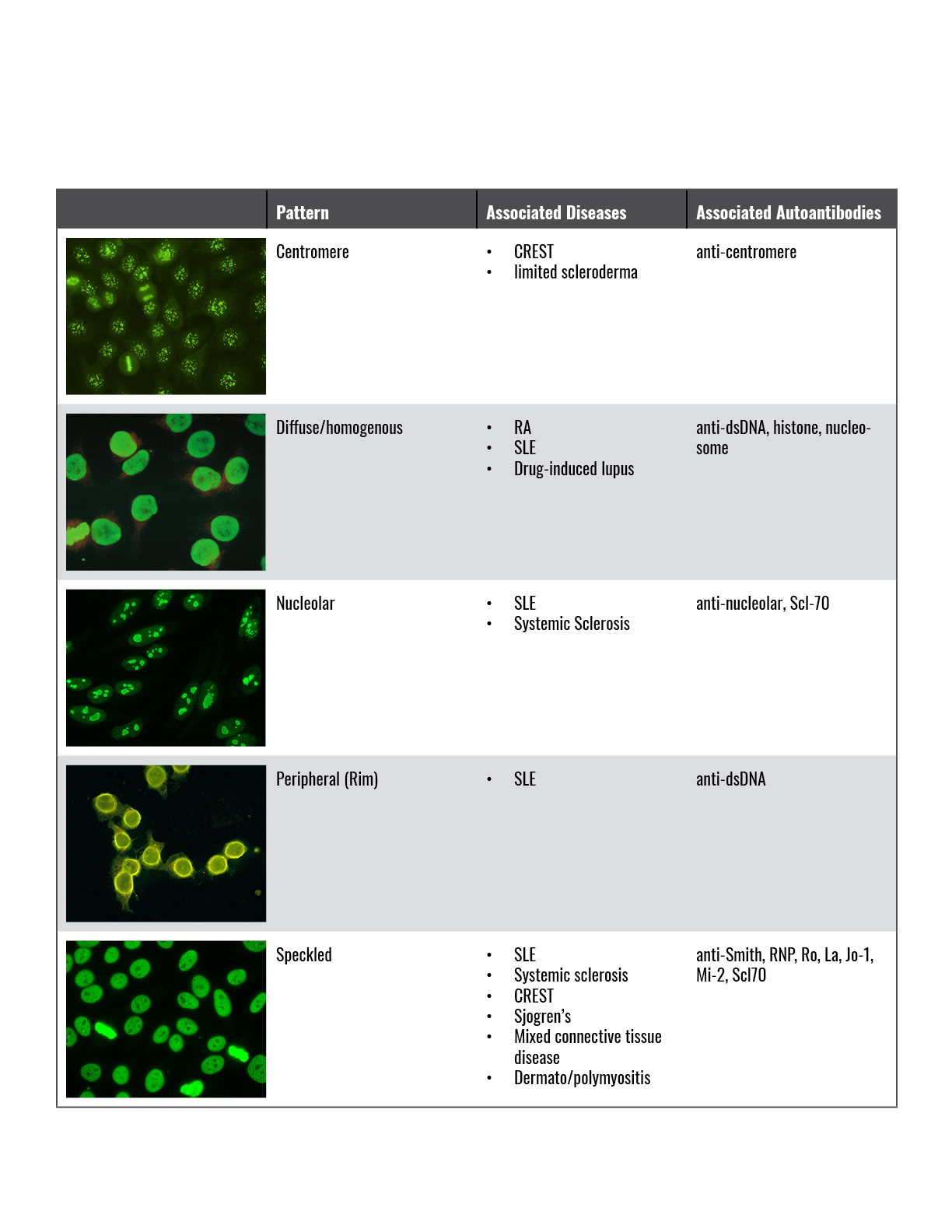
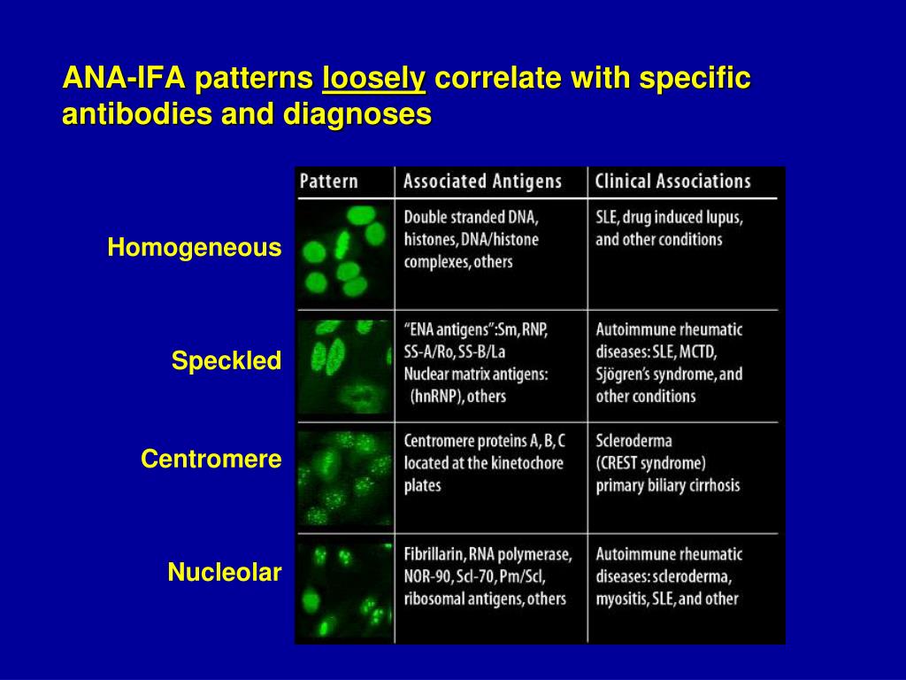
Homogeneous Ana Patterns - Web the most frequently observed ana patterns were the speckled (52.1%) and homogeneous (35.2%) patterns, while other patterns were rare representing less than 7% of the patients each. This is the most common pattern and can be seen with any autoimmune disease. If the test finds antinuclear antibodies in your blood, it may mean you have an autoimmune disorder. Web ana test results are most often reported in 2 parts: A homogenous staining pattern means the entire nucleus is stained with ana. Web a homogeneous/peripheral pattern reflects antibodies to histone/dsdna/chromatin, whereas many other specificities found in systemic rheumatic diseases show speckled patterns of various sizes and densities (fine speckled, large speckled, etc.). Web a homogenous (diffuse) pattern appears as total nuclear fluorescence and is common in people with systemic lupus. An ana test looks for antinuclear antibodies in your blood. Medically reviewed by scott zashin, md. Web her ana titer is 1:80, with a homogenous pattern.

DFS70 antibodies biomarkers for the exclusion of ANAassociated
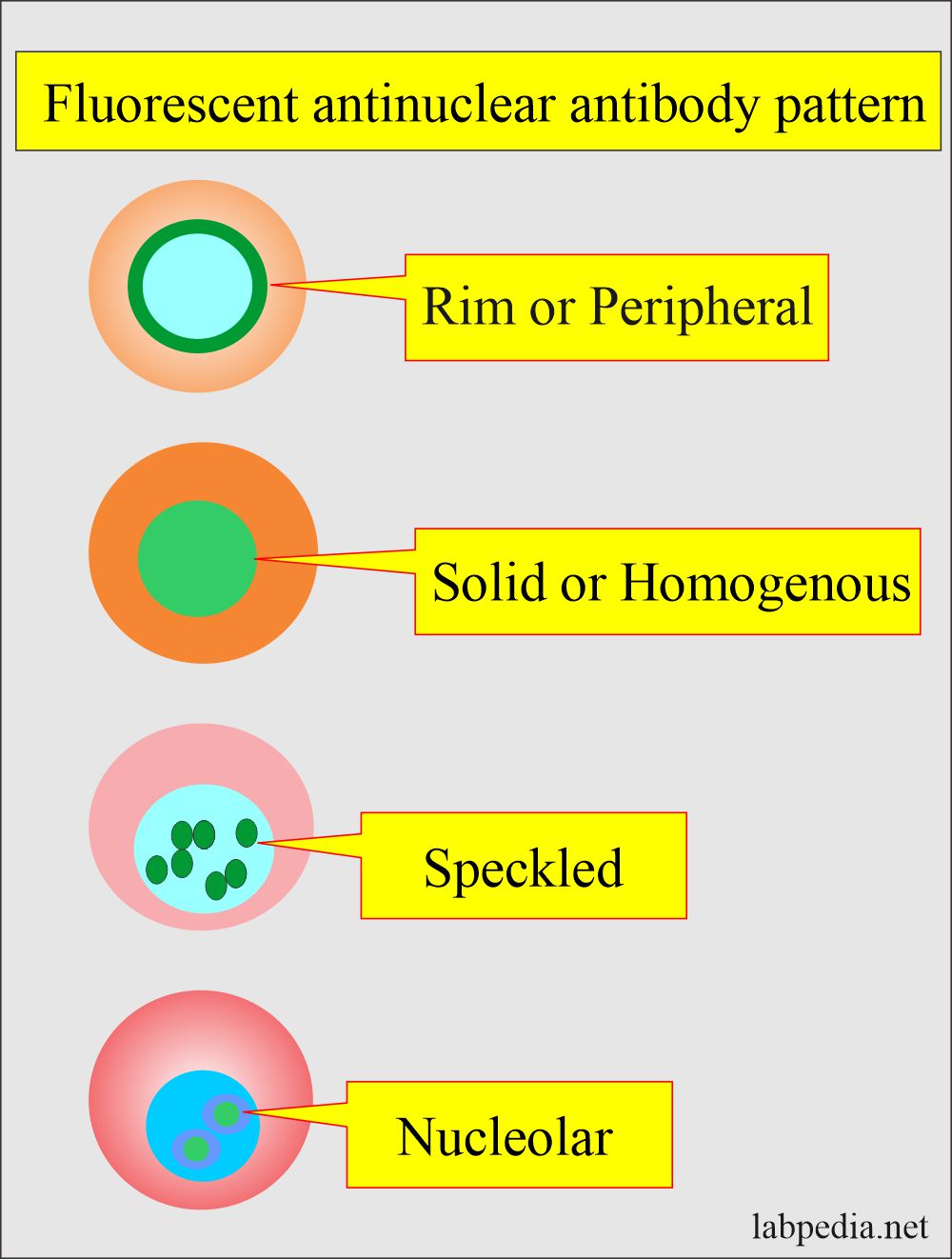
Web her ana titer is 1:80, with a homogenous pattern. The entire nucleus is stained with ana. A speckled staining pattern means fine, coarse speckles.

Common ANA patterns by IIF a, negative sample; b, homogeneous; c
The nucleoli maybe stained or not stained depending on cell substrate. Web antibodies that attack healthy proteins within the cell nucleus are called antinuclear antibodies.
Ana Test Patterns
A homogenous pattern can mean any autoimmune disease but more specifically, lupus or sjögren’s syndrome. Web what is an ana (antinuclear antibody) test? Homogenous fluorescence.

ANA Patterns
This is the most common pattern and can be seen with any autoimmune disease. Homogeneous and regular fluorescence across all nucleoplasm. A peripheral pattern indicates.

6. IFA pattern Homogeneous ANA pattern YouTube
This pattern is almost exclusive to systemic lupus. A titer (a measure of how much ana is in the blood) and a pattern (where the.
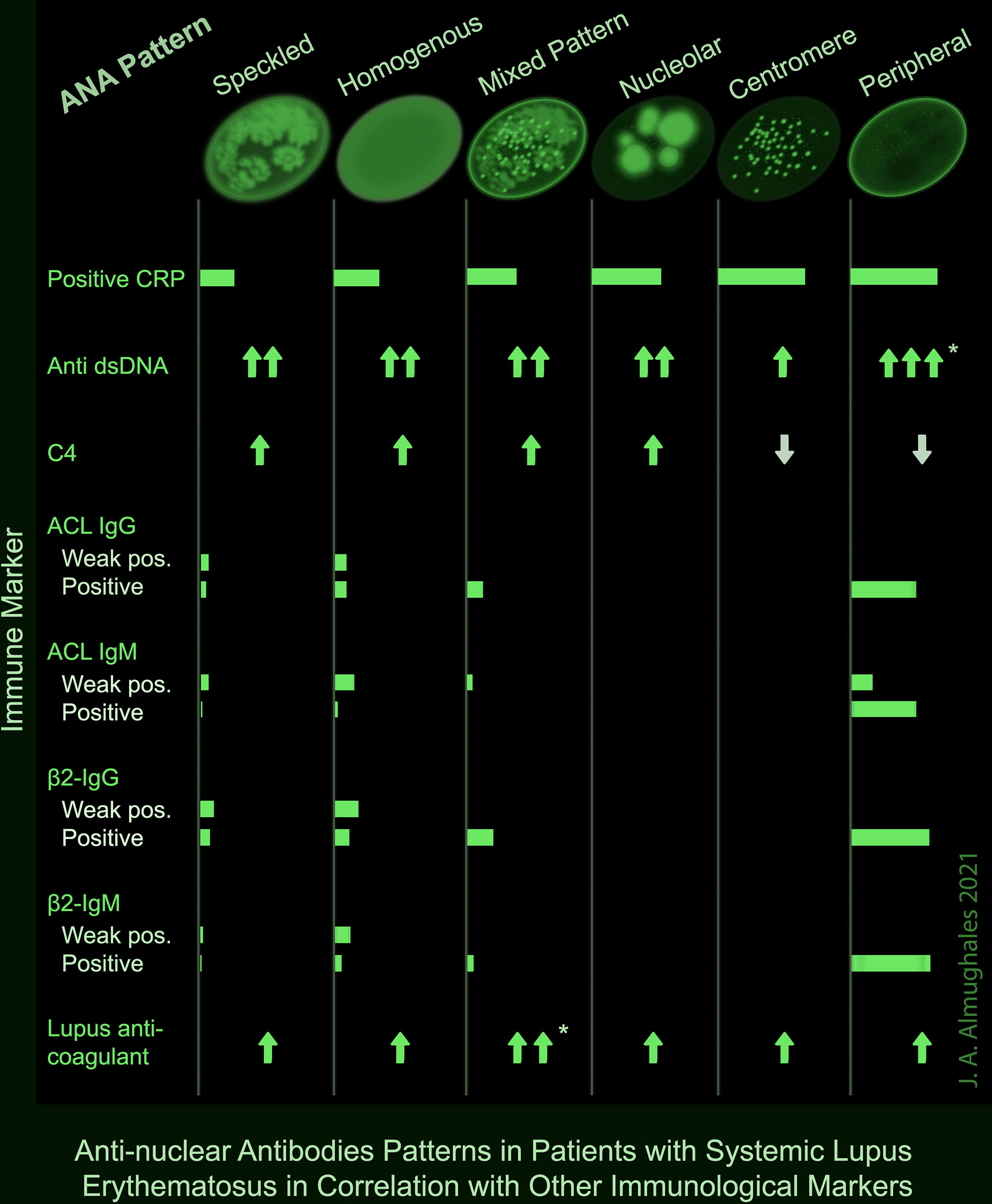
Frontiers AntiNuclear Antibodies Patterns in Patients With Systemic
Mitotic cells (metaphase, anaphase, and telophase) have the chromatin mass intensely stained in a homogeneous hyaline fashion. Web welcome to anapatterns.org, the official website for.
Antinuclear Factor (ANF), Antinuclear Antibody (ANA) and Its
The patient in case 1 has no end organ involvement of her lupus, yet her ana titer is > 1:1280. Web antibodies that attack healthy.

ANA Patterns
Titres are reported in ratios, most often 1:40, 1:80, 1:160, 1:320, and 1:640. Homogeneous — staining is even in the entire nucleus and is commonly.
.jpg)
ANA Mixed pattern University of Birmingham
Web ana test results are most often reported in 2 parts: An ana test detects antinuclear antibodies (ana) in your blood. If the test finds.
Homogeneous Ana Pattern Pagswa
A speckled pattern is also found in lupus. This pattern is almost exclusive to systemic lupus. Web each pattern is assigned an alphanumeric ac code.
A Membranous Pattern May Show Antibodies To Membrane Proteins.
This pattern is more commonly associated with antibodies. The patient in case 1 has no end organ involvement of her lupus, yet her ana titer is > 1:1280. Web a homogeneous/peripheral pattern reflects antibodies to histone/dsdna/chromatin, whereas many other specificities found in systemic rheumatic diseases show speckled patterns of various sizes and densities (fine speckled, large speckled, etc.). An ana test looks for antinuclear antibodies in your blood.
A Speckled Staining Pattern Means Fine, Coarse Speckles Of Ana Are Present.
Some, but not all labs will report a titre above 1:160 as positive. Web the presence of ana with a homogeneous & speckled (hs) pattern was significantly associated with the absence of cancer ( < 0.01). The entire nucleus is stained with ana. Web by carol eustice.
A Titer (A Measure Of How Much Ana Is In The Blood) And A Pattern (Where The Ana Was Detected In The Cells).
Homogeneous — staining is even in the entire nucleus and is commonly found in people with sle and discoid lupus erythematosus (dle). How the test is performed. Titres are reported in ratios, most often 1:40, 1:80, 1:160, 1:320, and 1:640. Doctors may order an ana test if you have signs or symptoms of an autoimmune disorder.
It Is Cases Such As These That Have Piqued The Interest Of Rheumatologists In The Ana And Its Role In Sle.
Interphase cells show homogeneous nuclear staining while mitotic cells show staining of the condensed chromosome regions. Medically reviewed by scott zashin, md. Web antibodies that attack healthy proteins within the cell nucleus are called antinuclear antibodies (anas). Fine and coarse speckles of ana staining are seen throughout the nucleus.