Fluorescein Staining Patterns - Web how to interpret fluorescein staining patterns to assist in red eye diagnosis. , chie sotozono a, kazuo tsubota d. 8.4mm oz observe apical clearance and insufficient peripheral clearance It is also useful as a. Web to assess total ocular surface staining, the cornea and conjunctival staining are graded, first using fluorescein dye to assess tear film stability over the cornea and then lissamine green or rose bengal dyes for conjunctival staining. Web in these cases, the staining pattern of fluorescein, which appears as bright green punctate lesions on the cornea, is evaluated and given a score. 4.3 (3 reviews) superficial punctate keratitis (spk) click the card to flip 👆. The fluorescein is applied on a moistened sterile strip of paper to the inner lining of the lower eyelid or applied in an eye drop mixed with a topical anesthetic. Web fluorescein penetrates poorly into the lipid layer of the corneal epithelium, and therefore, it does not stain normal cornea. Web corneal staining is often part of a routine eye exam.
Fluorescein Sodium Strips and Fluorescein angiography Eye Health Nepal
However, grading of des severity exactly by carefully assessing these dots is a rather difficult task for humans. Web fluorescein penetrates poorly into the lipid.

Classification of Fluorescein Breakup Patterns A Novel Method of
Isolated or diffuse dots across a larger area of the cornea. It is also useful as a. Web fluorescent staining is a pivotal molecular technique.
Corneal Manifestations of Systemic Diseases
4.3 (3 reviews) superficial punctate keratitis (spk) click the card to flip 👆. Web fluorescein penetrates poorly into the lipid layer of the corneal epithelium,.

Schematic representation of various patterns of corneal fluorescein
(a) dendritiform staining (dichotomously branching lesions, often with terminally bulbous swellings)—typical of herpes simplex keratitis. Web fluorescein staining patterns flashcards | quizlet. Web after fluorescein.

Classification of fluorescein breakup patterns. Download Scientific
20 these flecks and small stains are termed punctate epithelial erosions when focal defects are noted, or superficial punctate keratiti. When antibiotic treatment is indicated.
PPT Fluorescein Patterns PowerPoint Presentation, free download ID
8.4mm oz observe apical clearance and insufficient peripheral clearance (a) dendritiform staining (dichotomously branching lesions, often with terminally bulbous swellings)—typical of herpes simplex keratitis. Web.

Fluorescein Staining Patterns
Web how to interpret fluorescein staining patterns to assist in red eye diagnosis. This method involves fluorescent dyes or proteins that absorb light at a.

Figure 6 from Assessment of Corneal Fluorescein Staining in Different
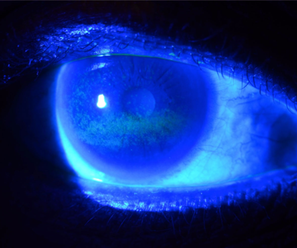
Web after fluorescein staining of the cornea, an abrasion will appear yellow under normal light and green in cobalt blue light. Here, we investigated the.

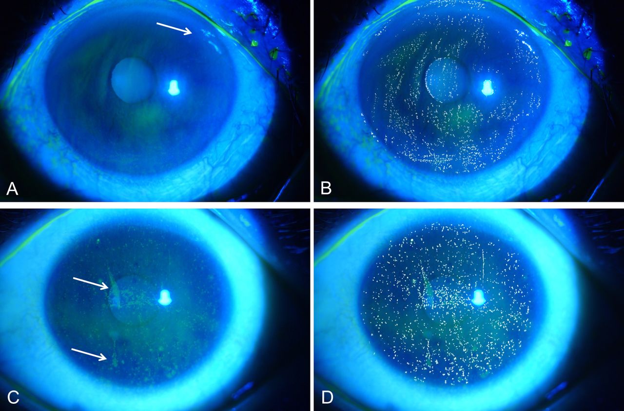
Clinical grading of fluorescein staining patterns, a grade 1, b grade
Here, we investigated the differences in the clinical features of ded patients with and without pp corneal staining (ppcs). Web corneal staining is often part.

Diagnostics Free FullText Characteristics and Utility of
Isolated or diffuse dots across a larger area of the cornea. Web fluorescent staining is a pivotal molecular technique that brings to light the structures.
Web After Fluorescein Staining Of The Cornea, An Abrasion Will Appear Yellow Under Normal Light And Green In Cobalt Blue Light.
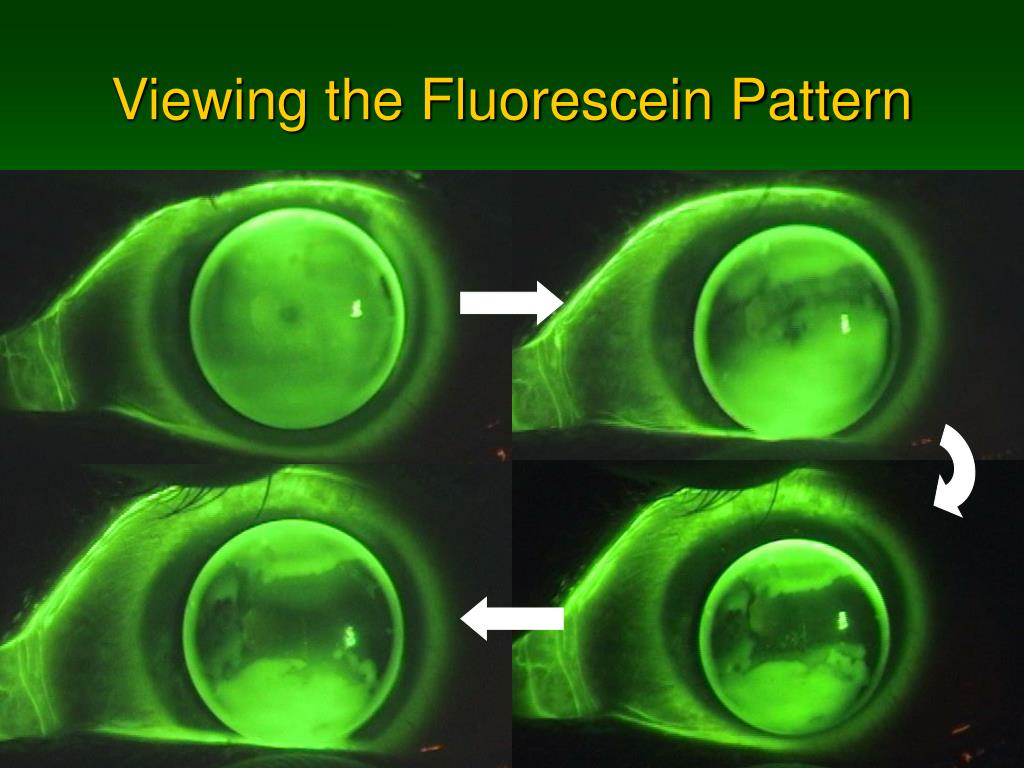
Web to assess total ocular surface staining, the cornea and conjunctival staining are graded, first using fluorescein dye to assess tear film stability over the cornea and then lissamine green or rose bengal dyes for conjunctival staining. 20 these flecks and small stains are termed punctate epithelial erosions when focal defects are noted, or superficial punctate keratiti. Web how to interpret fluorescein staining patterns to assist in red eye diagnosis. Notice greater fluorescence on k bc with a 9.5mm dia;
8.4Mm Oz Observe Apical Clearance And Insufficient Peripheral Clearance
Physicians should carefully examine for foreign bodies and remove. Web the application of fluorescein also extends to bioimaging whole anatomic structures and even further to cellular components in immunohistological staining. Isolated or diffuse dots across a larger area of the cornea. Web fluorescein penetrates poorly into the lipid layer of the corneal epithelium, and therefore, it does not stain normal cornea.
This Article Outlines The Indications, Mechanism Of Action, Adverse Event Profile, And Contraindications For Fluorescein To Guide The Healthcare Team In Evaluating Ophthalmic.
The ocular surface is most commonly stained with fluorescein dye for dry eye assessment. Similarly, the proper use and diagnostic. Web fluorescent staining is a pivotal molecular technique that brings to light the structures within biological cells and tissues through fluorescence. Web in these cases, the staining pattern of fluorescein, which appears as bright green punctate lesions on the cornea, is evaluated and given a score.
However, Grading Of Des Severity Exactly By Carefully Assessing These Dots Is A Rather Difficult Task For Humans.
Web the most common type is superficial punctate staining, which manifests as patterns of small dots or flecks that stain brightly with fluorescein in either a diffuse pattern or segregated patches on certain areas. It aids in the diagnosis of various ocular pathologies. When antibiotic treatment is indicated in various eye conditions. Dry eye disease, fluorescein, interpretation, ocular surface staining, vital dyes.