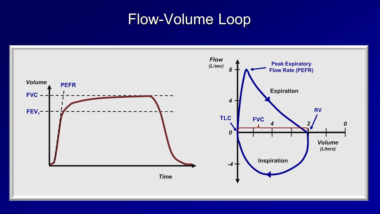
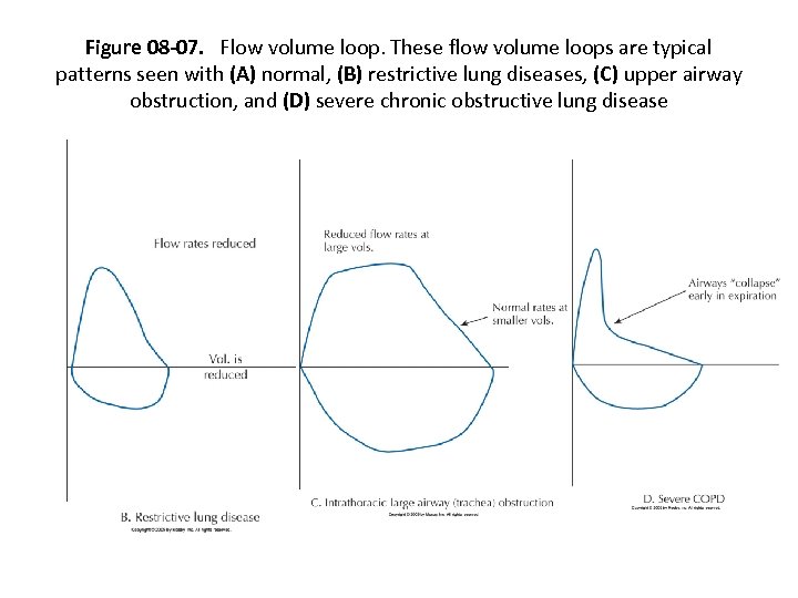
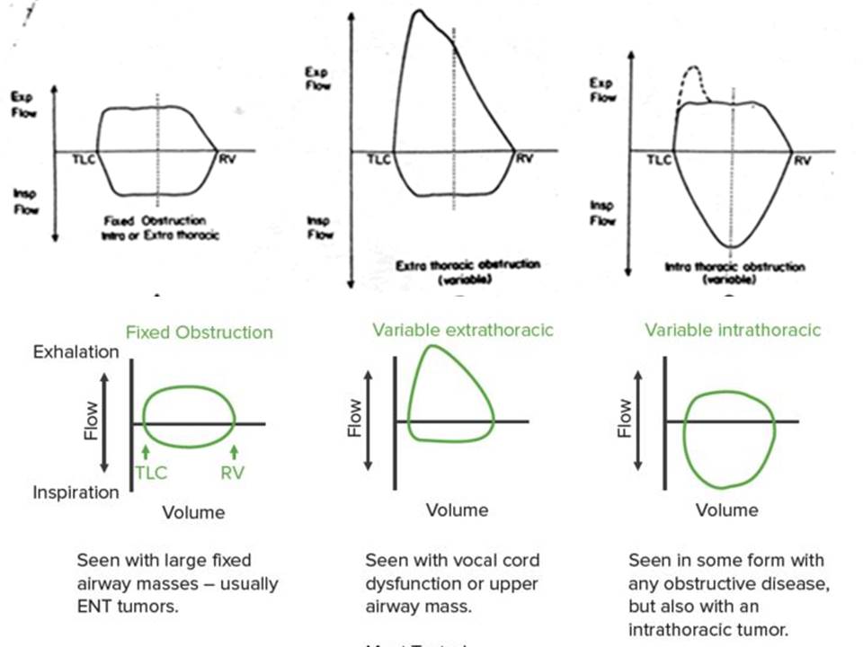
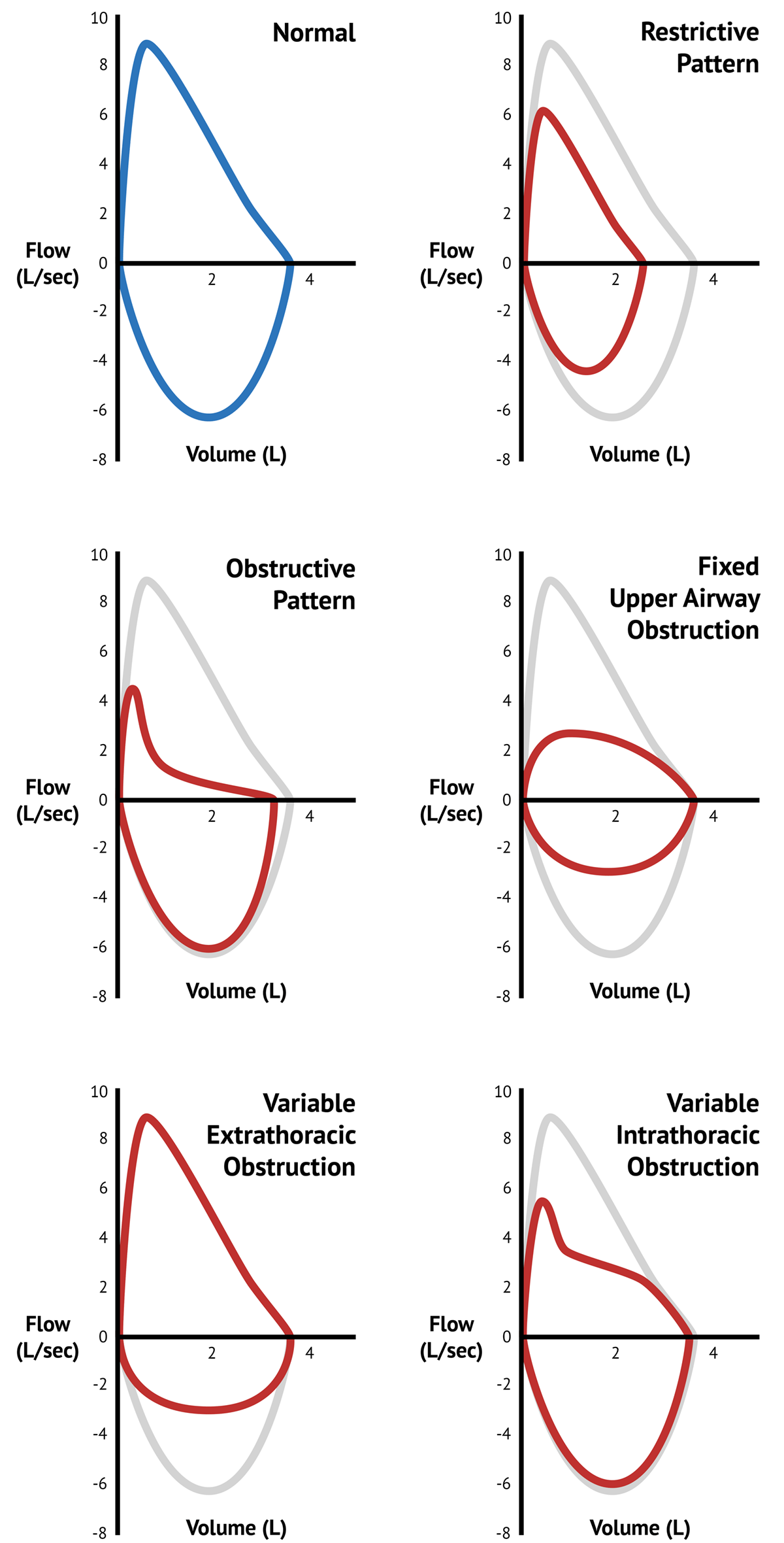
Flow Volume Loop Patterns - Changes in the contour of the loop can aid in the diagnosis and localization of airway obstruction [ 1 ]. Web pattern recognition is key.a low fev1/fvc ratio (the forced expiratory volume in 1 second divided by the forced vital capacity) indicates an obstructive pattern, whereas a normal value indicates either a restrictive or a normal pattern. Thumbnail versions are inadequate for this purpose and should be avoided. After the pef the curve descends (=the flow decreases) as more air is expired. Web airflow and lung volume measurements can be used to differentiate obstructive from restrictive pulmonary disorders, to characterize severity, and to measure responses to therapy. This finding has also been described as “two compartments.” a ct chest was obtained which showed an obstructing right mainstem bronchus lesion. Thorough understanding of both scalars and loops, and their characteristic appearances, is essential to being able to evaluate a patient’s respiratory mechanics and interaction with the. Neuromuscular weakness #diagnosis #pfts #spirometry #flowvolume. Web flow volume loops. Low maximum forced expiratory flow, biphasic expiratory curve, flow oscillations, and notching.
Pulmonary Function Tests (PFT) Lesson 2 Spirometry YouTube
Low maximum forced expiratory flow, biphasic expiratory curve, flow oscillations, and notching. This finding has also been described as “two compartments.” a ct chest was.
Pulmonary Function Testing Chapter 8 Pulmonary Function
Low maximum forced expiratory flow, biphasic expiratory curve, flow oscillations, and notching. Airflow and lung volume measurements can be used to differentiate obstructive from restrictive.

Pulmonary Function Testing Flow Volume Loop Patterns GrepMed
Estimates of the number of individuals with flow oscillation range from 1.4% to 13.4% with the higher estimates being observed primarily with inspiratory loops. Low.

PPT Pulmonary Function Tests PowerPoint Presentation, free download
Airflow and lung volume measurements can be used to differentiate obstructive from restrictive pulmonary disorders, to characterize severity, and to measure responses to therapy. Neuromuscular.
Flow Volume Loops in Spirometry
Restrictive parenchymal lung disease h. Unilateral main stem bronchial obstruction d. After the pef the curve descends (=the flow decreases) as more air is expired..

Flow Volume Loops Respiratory Physiology Pulmonary Medicine YouTube
At the start of the test both flow and volume are equal to zero. Low maximum forced expiratory flow, biphasic expiratory curve, flow oscillations, and.

Pulmonary FlowVolume Loops & Disease Medical science, Pathology
The test is easy to demonstrate, administer, and analyze. Web pattern recognition is key.a low fev1/fvc ratio (the forced expiratory volume in 1 second divided.
FlowVolume Loops Lung Function Tests MedSchool
Provide a graphical analysis of inspiratory and expiratory flow from various inspired lung volumes. Fev 3, forced expiratory volume in the third second; Web flow.
PPT Pulmonary Function Testing PowerPoint Presentation, free download
Thumbnail versions are inadequate for this purpose and should be avoided. The airways are divided into intrathoracic and extrathoracic components by the thoracic inlet. Low.

Pulmonary Function Tests Pulmonary Medbullets Step 2/3
At the start of the test both flow and volume are equal to zero. The test is easy to demonstrate, administer, and analyze. Failure of.
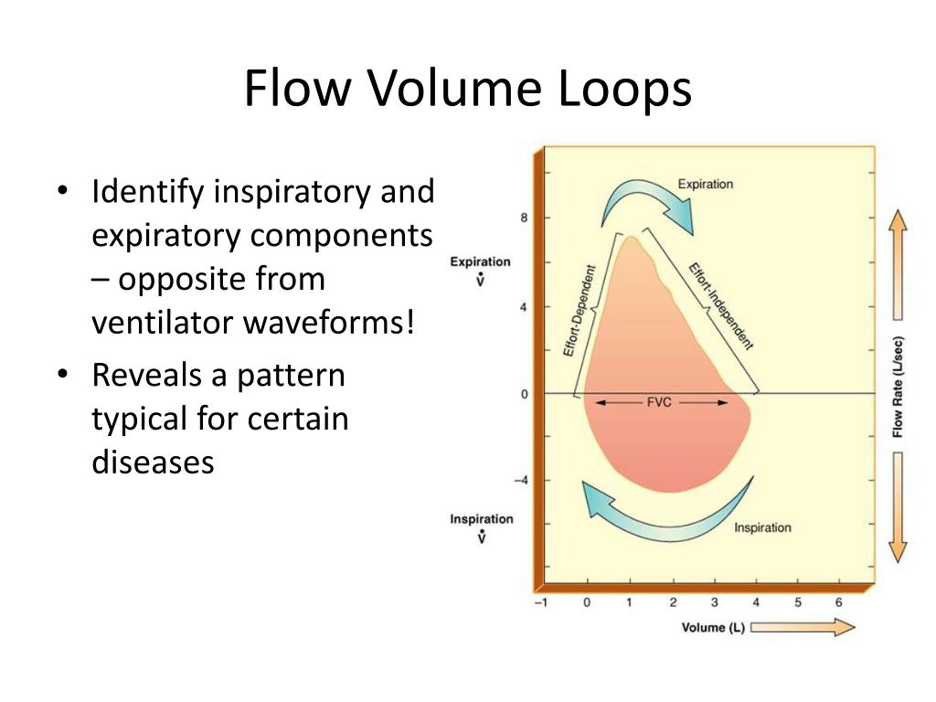
Web Flow Volume Loops.
Web in summary, we suggest that this flow volume loop pattern observed in our patient is diagnostic of mainstem endobronchial tumors which cause nearly complete obstruction during forced exhalation maneuvers. Fev 1, forced expiratory volume in the first second; The test is easy to demonstrate, administer, and analyze. After the starting point the curve rapidly mounts to a peak:
Low Maximum Forced Expiratory Flow, Biphasic Expiratory Curve, Flow Oscillations, And Notching.
Thumbnail versions are inadequate for this purpose and should be avoided. Failure of the expiratory flow curve to reach zero (an open loop) a scooped out expiratory flow pattern. Provide a graphical analysis of inspiratory and expiratory flow from various inspired lung volumes. This case is similar to one reported by dull and coworkers.
Airflow And Lung Volume Measurements Can Be Used To Differentiate Obstructive From Restrictive Pulmonary Disorders, To Characterize Severity, And To Measure Responses To Therapy.
Fixed upper airway obstruction (uao) e. Fev 3, forced expiratory volume in the third second; Low maximum forced expiratory flow, biphasic expiratory curve, flow oscillations, and notching. Web airflow and lung volume measurements can be used to differentiate obstructive from restrictive pulmonary disorders, to characterize severity, and to measure responses to therapy.
After The Pef The Curve Descends (=The Flow Decreases) As More Air Is Expired.
Neuromuscular weakness #diagnosis #pfts #spirometry #flowvolume. This finding has also been described as “two compartments.” a ct chest was obtained which showed an obstructing right mainstem bronchus lesion. The sawtooth pattern occurs in only a small fraction of patients but it is quite noticeable when you see it. Restrictive parenchymal lung disease h.