Fibrillar Pattern Acral Nevus - 3.2 diffuse pigmentation of variable shades of brown. Web alm ( acral lentiginous melanoma ), am ( acral melanoma ), amf ( atypical melanoma of foot ), cm ( cutaneous melanoma ), fish ( fluorescent in situ hybridization. Web acral melanocytic nevi have three major dermoscopic patterns: Web acral lentiginous melanoma (alm), first described by reed in 1976, is a histological subtype of cutaneous melanoma arising on the acral areas 1, 2. Web acral melanocytic nevus is characterized by a parallel furrow pattern, whereas acral melanoma has a parallel ridge pattern. Web dermoscopy of melanoma according to different body sites: Web we conclude that the double line variant of the parallel furrow pattern and crista dotted pattern, which probably correspond to the congenital type acral nevus,. Web unfortunately, the furrow pattern is not the only pattern seen in acral nevi. Other dermatoscopic patterns have descriptive names, such as latticelike, fibrillar,. Acral melanocytic nevi have three major dermoscopic patterns:

Schematic illustration of the observed acral benign dermoscopic
Web acral melanocytic nevus is characterized by a parallel furrow pattern, whereas acral melanoma has a parallel ridge pattern. 3.2 diffuse pigmentation of variable shades.

Dermoscopic Patterns of Acral Melanocytic Nevi Their Variations
Web unfortunately, the furrow pattern is not the only pattern seen in acral nevi. Web acral lentiginous melanoma (alm), first described by reed in 1976,.
Acral Nevus (ICD10 D22) Online consultation AI dermatologist
Web acral melanocytic lesion displaying fibrillar pattern on dermoscopy. Most individuals have a predominant type of. Web acral lentiginous melanoma (alm), first described by reed.
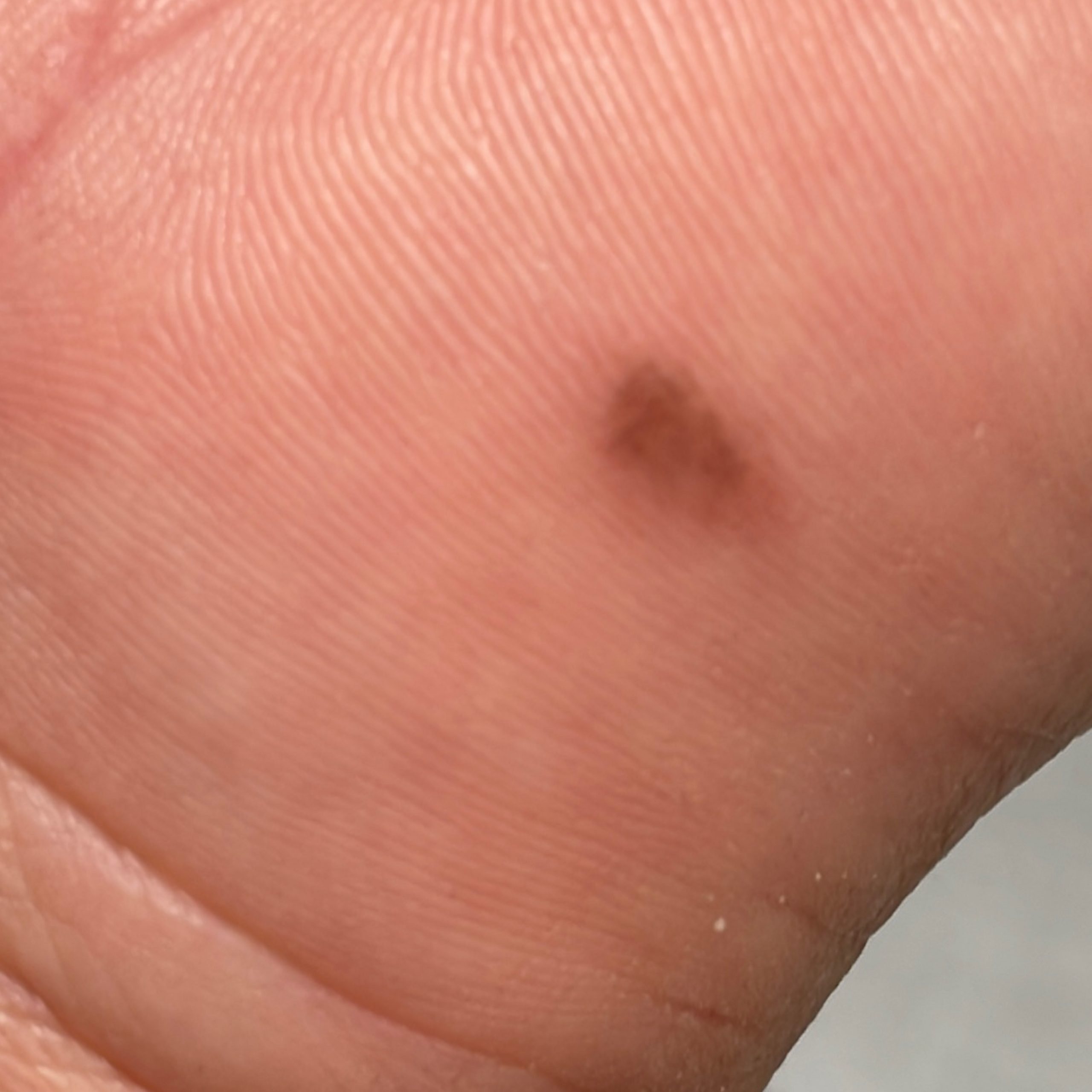
acral pictures, photos
The fibrillar pattern is a benign dermoscopic pattern that exhibits fine fibrillar or filamentous pigmented lines that run obliquely to the skin markings. Web dermoscopy.

Variations in the Dermoscopic Features of Acquired Acral Melanocytic
3.2 diffuse pigmentation of variable shades of brown. Web acral melanocytic lesion displaying fibrillar pattern on dermoscopy. Web acral melanocytic nevi have three major dermoscopic.

Regular fibrillar pattern of acral nevus (dermoscopy with the furrow
Web alm ( acral lentiginous melanoma ), am ( acral melanoma ), amf ( atypical melanoma of foot ), cm ( cutaneous melanoma ), fish.

Dermatopathology Free FullText Acral Melanocytic Neoplasms A
Web acral lentiginous melanoma (alm), first described by reed in 1976, is a histological subtype of cutaneous melanoma arising on the acral areas 1, 2..

Dermoscopy Made Simple Acral nevus
Web we conclude that the double line variant of the parallel furrow pattern and crista dotted pattern, which probably correspond to the congenital type acral.

Dermoscopic Changes in Acral Melanocytic Nevi During Digital Followup
Most individuals have a predominant type of. Web unfortunately, the furrow pattern is not the only pattern seen in acral nevi. Acral melanocytic nevi have.

dermoscopy Fibrillar pattern
Web unfortunately, the furrow pattern is not the only pattern seen in acral nevi. Web 2.3 fibrillar pattern. Acral melanocytic nevi have three major dermoscopic.
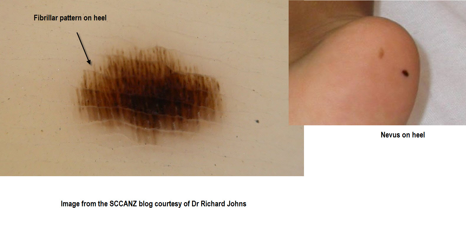
Web By Pressing The Dermatoscope Against A Lesion While Moving The Dermatoscope Horizontally On The Skin, A Parallel Furrow Pattern May Be Transformed Into A Fibrillar Pattern (Or Vice.
Most individuals have a predominant type of. Web alm ( acral lentiginous melanoma ), am ( acral melanoma ), amf ( atypical melanoma of foot ), cm ( cutaneous melanoma ), fish ( fluorescent in situ hybridization. Web dermoscopy of melanoma according to different body sites: Web unfortunately, the furrow pattern is not the only pattern seen in acral nevi.
Acral Melanocytic Nevi Have Three Major Dermoscopic Patterns:
Web we conclude that the double line variant of the parallel furrow pattern and crista dotted pattern, which probably correspond to the congenital type acral nevus,. 3.2 diffuse pigmentation of variable shades of brown. Other dermatoscopic patterns have descriptive names, such as latticelike, fibrillar,. Web 2.3 fibrillar pattern.
Pigmentation Following And Crossing The Furrows In Acral Melanocytic Nevi Fibrillar.
Web in contrast, the main dermoscopic pattern of acral nevus is the parallel furrow pattern (pfp) showing linear pigmentation along the sulci of the skin markings. Web acral lentiginous melanoma (alm), first described by reed in 1976, is a histological subtype of cutaneous melanoma arising on the acral areas 1, 2. The fibrillar pattern is a benign dermoscopic pattern that exhibits fine fibrillar or filamentous pigmented lines that run obliquely to the skin markings. Web acral melanocytic nevi have three major dermoscopic patterns:
Web Acral Melanocytic Lesion Displaying Fibrillar Pattern On Dermoscopy.
Web acral melanocytic nevus is characterized by a parallel furrow pattern, whereas acral melanoma has a parallel ridge pattern.