Ekg Patterns - Web what is an ecg? The t wave represents the recovery phase of the ventricles. Are you learning to interpret ecgs? Electrodes are placed on different parts of a patient’s limbs and chest to record the electrical activity. V5, v6 = l side of the heart. Litfl ecg library is a free educational resource covering over 100 ecg topics relevant to emergency medicine and critical care. Osborne wave (j wave) delta wave. An electrocardiogram, also called an ecg or ekg, is a simple and painless test that measures the electrical impulses of your heart. The heart's rhythm is the time between each heartbeat. For more medicine videos consider subscrib.
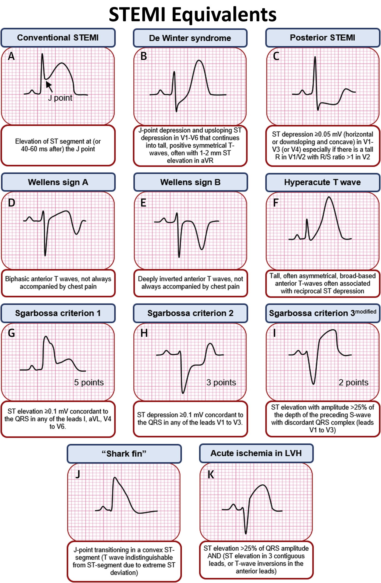
STEMI Equivalents on ECG • Conventional STEMI Elevation GrepMed
It is used to record the electrical activity of the heart from different angles to both identify and locate pathology. The image is credited to.

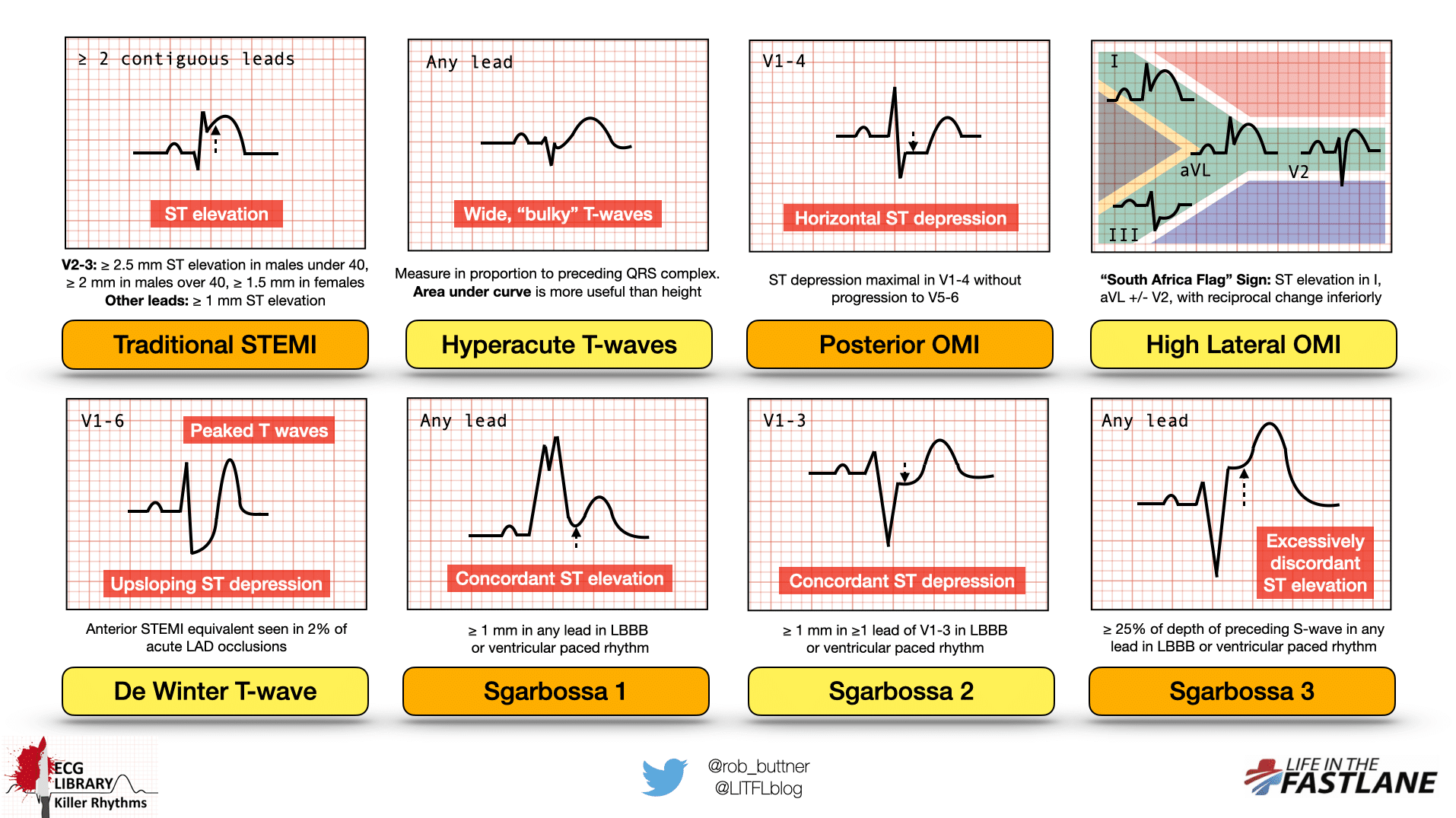
11 ECG patterns strongly suggestive of GrepMed
If a patient has a regular heart rhythm, their heart rate can be calculated using the following method: Therefore, ecg interpretation requires a structured assessment.

How to read a normal ECG(Electrocardiogram)? HubPages
Web the electrical activity on an ecg (ekg). An electrocardiogram — abbreviated as ekg or ecg — measures the electrical activity of the heartbeat. These.

Understanding the EKG Signal Atrial Fibrillation Resources for Patients
Describe cardiac anatomy and physiology. Web the ecg must always be interpreted systematically. Hyperacute (peaked) t waves or pseudonormalisation of previously inverted t waves (i.e..

ECG interpretation Characteristics of the normal ECG (Pwave, QRS
The areas represented on the ecg are summarized below: If a patient has a regular heart rhythm, their heart rate can be calculated using the.
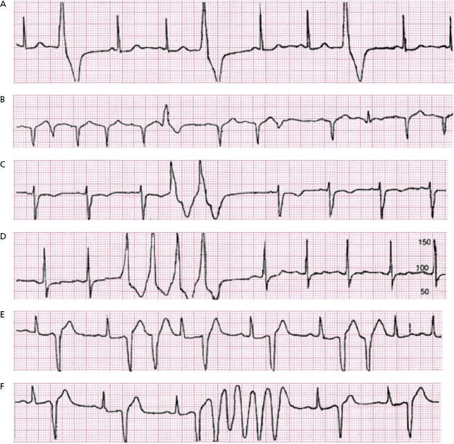
12 ECG Patterns of Ventricular Arrhythmias Thoracic Key
Hyperacute (peaked) t waves or pseudonormalisation of previously inverted t waves (i.e. The interpretation algorithm presented below is easy to follow and it can be.
Killer ECG Patterns Part 2 • LITFL • ECG Library
The t wave represents the recovery phase of the ventricles. Web other ecg patterns of ischaemia. Web what is an ecg? Becoming upright) suggest hyperacute.

RCAT for Arrhythmias EKG Pocket Reference Guide Plus EKG Badge Great
An electrocardiogram — abbreviated as ekg or ecg — measures the electrical activity of the heartbeat. An ecg can show irregular heartbeats, called arrhythmias. An.
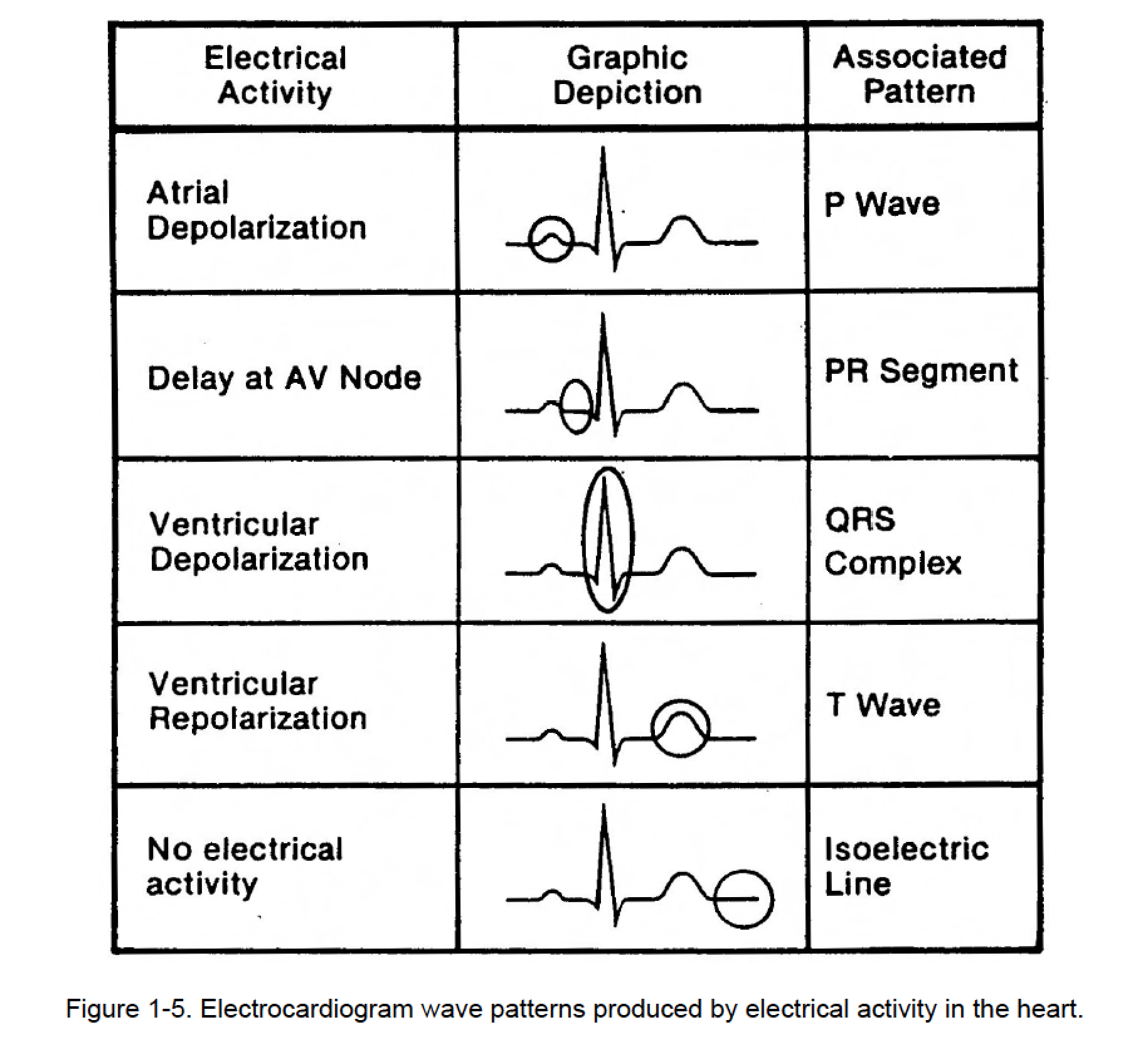
Figure 15. Cardiac Rhythm Interpretation
Web the ecg must always be interpreted systematically. Web typical ecg patterns you should be familiar with. We look at the most common ecg rhythms.

practice ekg rhythm strips for acls
We look at the most common ecg rhythms and patterns seen in medicine, including main identifying features of each. Web continuing education activity. Web 6.
The P Wave Represents The Electrical Impulse Traveling Through The Atria, While The Qrs Complex Indicates The Impulse Moving Through The Ventricles.
These patients require immediate cardiology referral for emergent reperfusion therapy. A medical provider’s ability to correctly interpret and react to an ecg could mean the difference between life and death for a patient. Web typical ecg patterns you should be familiar with. Web overview of the normal electrocardiogram (ecg) ecg interpretation includes an assessment of the morphology (appearance) of the waves and intervals on the ecg curve.
One Of The Most Useful And Commonly Used Diagnostic Tools Is Electrocardiography (Ekg) Which Measures The.
An electrocardiogram, also called an ecg or ekg, is a simple and painless test that measures the electrical impulses of your heart. Divide 300 by this number to calculate heart rate. Web a normal ecg pattern consists of different waves and intervals, each representing a specific part of the heart's electrical cycle. Becoming upright) suggest hyperacute stemi.
The Areas Represented On The Ecg Are Summarized Below:
We look at the most common ecg rhythms and patterns seen in medicine, including main identifying features of each. Ecg is the abbreviated term for an electrocardiogram. Apply leads for electrocardiograms (ecgs) and cardiac monitoring. With each beat, an electrical impulse (or “wave”) travels through the heart.
The Heart's Rhythm Is The Time Between Each Heartbeat.
Electrodes are placed on different parts of a patient’s limbs and chest to record the electrical activity. Web 6 min read. All our ecgs are free to reproduce for educational purposes, provided: Willem einthoven first invented it in 1902.