Ecg S1 S2 S3 Pattern - S1 s2 s3 pattern = far right axis deviation with dominant s waves in leads i, ii and iii. Table 4.2 ecg criteria for right. An amplitude of at least 1.5 mm — was found in 423 subjects (6.7%). Web abstract = the s1 s2 s3 pattern in the electrocardiogram has been variously defined. Web the purpose of this chapter is to review the role of the ecg in the diagnosis of cardiac chamber enlargement. St depression and t wave inversion in leads corresponding to the right ventricle: Web biatrial enlargement is diagnosed when criteria for both right and left atrial enlargement are present on the same ecg. There are 2 main heart sounds that can be heard during auscultation: Deep s waves in the left precordial leads v 5 and v 6 (r:s <1); Web in the limb leads right axis deviation develops and at times prominent q waves simulating an imi appear in leads 2,3, and avf.
Ecg criteria of chamber enlargement
Web in the limb leads right axis deviation develops and at times prominent q waves simulating an imi appear in leads 2,3, and avf. S1.
ECG Congenital Heart Disease
Web abstract = the s1 s2 s3 pattern in the electrocardiogram has been variously defined. The s1 and s2 heart sounds are part of the.
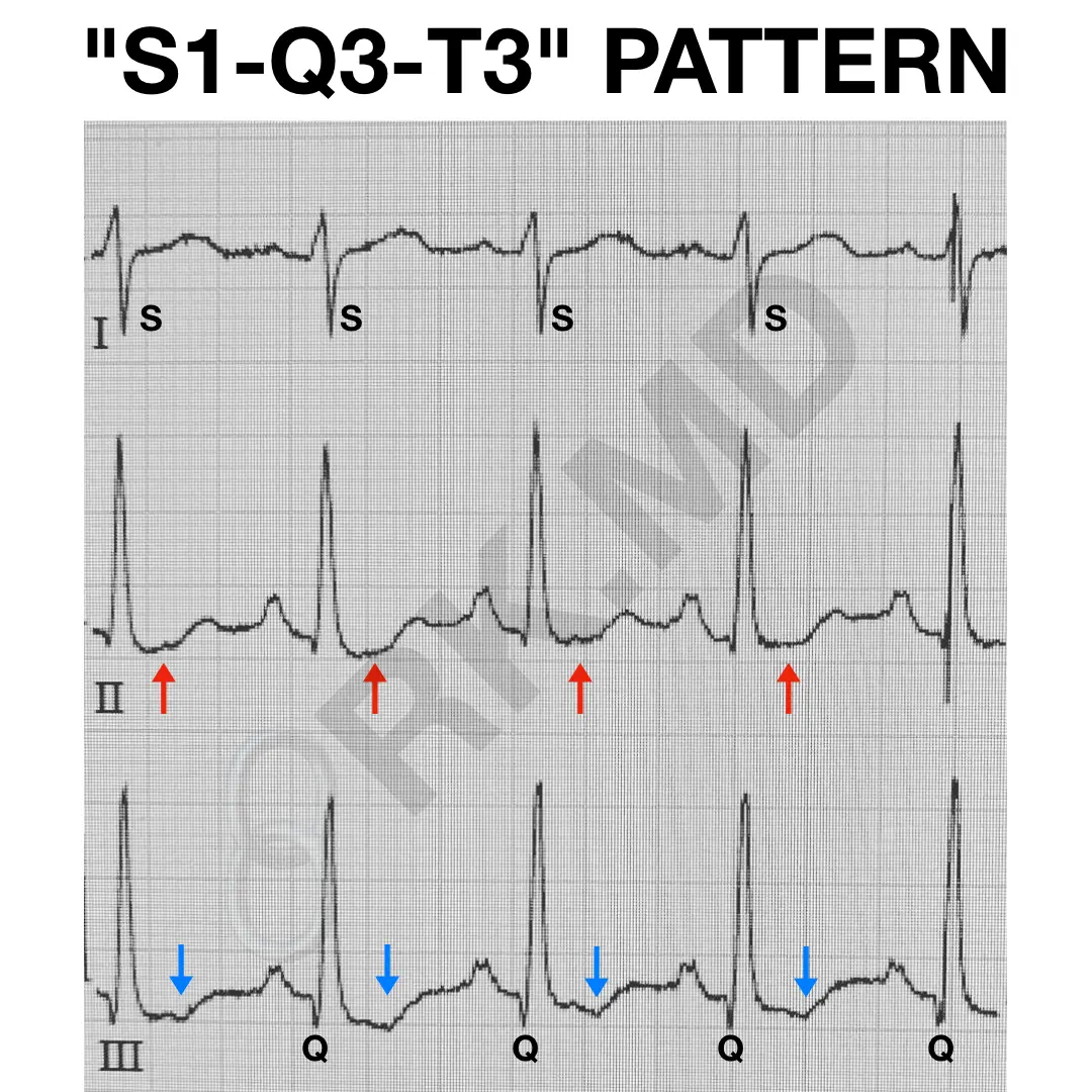
S1Q3T3 EKG Pattern RK.MD
S1 s2 s3 pattern = far right axis deviation with dominant s waves in leads i, ii and iii. Web in the limb leads right.
Figure 15. Cardiac Rhythm Interpretation
His electrocardiogram is an excellent example of the s1, s2, s3 syndrome. University of michigan murmur library. There are 2 main heart sounds that can.
Atlas of Electrocardiography Basicmedical Key
Some apply this term to all cases with an s wave in each standard lead, regardless of magnitude, while others use it to. Web in.

Standard (S1, S2, S3) and alternate (A1, A2, A3) ECG electrode
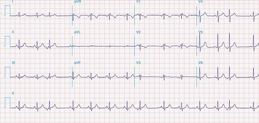
Note that there is a prominent s wave in leads 1, 2, and 3 and the s waves are equal in duration and magnitude to.

The Electrocardiogram explained What is an ECG?
Web the ecg changes associated with acute pulmonary embolism may be seen in any condition that causes acute pulmonary hypertension, including hypoxia causing pulmonary hypoxic.
Standard (S1, S2, S3) and alternate (A1, A2, A3) ECG electrode
The most typical ecg findings in emphysema are: An amplitude of at least 1.5 mm — was found in 423 subjects (6.7%). Web biatrial enlargement.

【コラム051】S1S2S3パターンを考えます。 Cardio2012のECGブログ
University of michigan murmur library. An amplitude of at least 1.5 mm — was found in 423 subjects (6.7%). There is a terminal r wave.

Description, criteria, and example of the different QRS morphologies
This latter application, which we prefer, is generally associated with marked. Web the s1 s2 s3 pattern in the electrocardiogram has been variously defined. An.
Ecg Criteria For Biatrial Enlargement.
Web ecg changes occur in chronic obstructive pulmonary disease (copd) due to: Web the purpose of this chapter is to review the role of the ecg in the diagnosis of cardiac chamber enlargement. An amplitude of at least 1.5 mm — was found in 423 subjects (6.7%). Web the ecg changes associated with acute pulmonary embolism may be seen in any condition that causes acute pulmonary hypertension, including hypoxia causing pulmonary hypoxic vasoconstriction.
The Diagnosis Of Biatrial Enlargement Requires Criteria For Lae And Rae To Be Met In Either Lead Ii, Lead V1 Or A Combination Of Leads.
Rv strain can be seen in leads v1 and v2 but also in leads 2,3, avf. The most typical ecg findings in emphysema are: Some apply this term to all cases with an s wave in each standard lead, regardless of magnitude, while others use it to. S1 heart sound corresponds to.
His Electrocardiogram Is An Excellent Example Of The S1, S2, S3 Syndrome.
Web the s1 s2 s3 pattern in the electrocardiogram has been variously defined. Web right ventricular strain is a repolarisation abnormality due to right ventricular hypertrophy (rvh) or dilatation. The s1 and s2 heart sounds are part of the normal heart sounds. An s wave deeper than r in all 3 standard leads) is a reliable index of rvh.
Web An S1, S2, S3 Pattern, Which May Mimic A Left Anterior Hemiblock, Is Frequently Associated With The Brugada Repolarization Abnormalities And Most Likely Reflects A Concomitant Intraventricular Conduction Defect Localized To The Right Ventricular Outflow Tract Due To The Disease Involvement Of The Peripheral Fascicles Of The Right Bundle Branch.
Inferior leads ii, iii, avf, often most pronounced in lead iii as this is the most rightward facing. S1 s2 s3 pattern = far right axis deviation with dominant s waves in leads i, ii and iii. Deep s waves in the left precordial leads v 5 and v 6 (r:s <1); St depression and t wave inversion in leads corresponding to the right ventricle: