Ecg Pulmonary Disease Pattern - Pulmonary pattern and what else? Electrocardiography (ecg) is a useful adjunct to other pulmonary tests because it provides information about the right side of. Web electrocardiographic (ecg) findings may help in clinical decision making regarding this disease entity. Associated with increased pulmonary artery pressures in the setting of acute or chronic right ventricular hypertrophy or dilatation: Web electrocardiographic (ecg) abnormalities associated with chronic obstructive pulmonary disease (copd) include right atrial enlargement, right. Our aim was to separate the effects on ecg by airway obstruction,. Web the ecg changes associated with acute pulmonary embolism may be seen in any condition that causes acute pulmonary hypertension, including hypoxia. Tetralogy of fallot, pulmonary stenosis) Web electrocardiography (ecg) in pulmonary disorders. Web a better understanding of the ecg changes in copd may improve interpretation of ecg in these patients and help revealing the dominant pathophysiology of their airway disease.

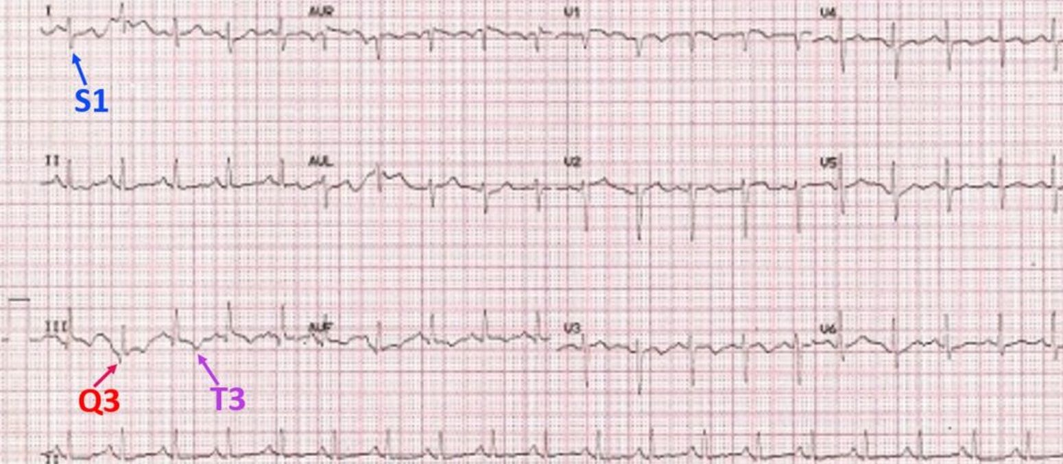
S1Q3T3 on ECG in a patient with Acute Pulmonary Embolism GrepMed
Associated with increased pulmonary artery pressures in the setting of acute or chronic right ventricular hypertrophy or dilatation: Pulmonary pattern and what else? Web electrocardiographic.
ECG in Chronic Obstructive Pulmonary Disease • LITFL • ECG Library
Web the following ecg signs reflecting ccp were collected: Web specific electrocardiographic abnormalities and cardiac arrhythmias are prevalent in chronic obstructive pulmonary disease. These changes.
Pulmonary embolism electrocardiogram wikidoc
The underlying pathophysiology is complex. Web ecg abnormalities are common in patients with pulmonary embolism, with the most frequent being sinus tachycardia, right ventricular strain,.
ECG Changes in Pulmonary Embolism New Health Advisor
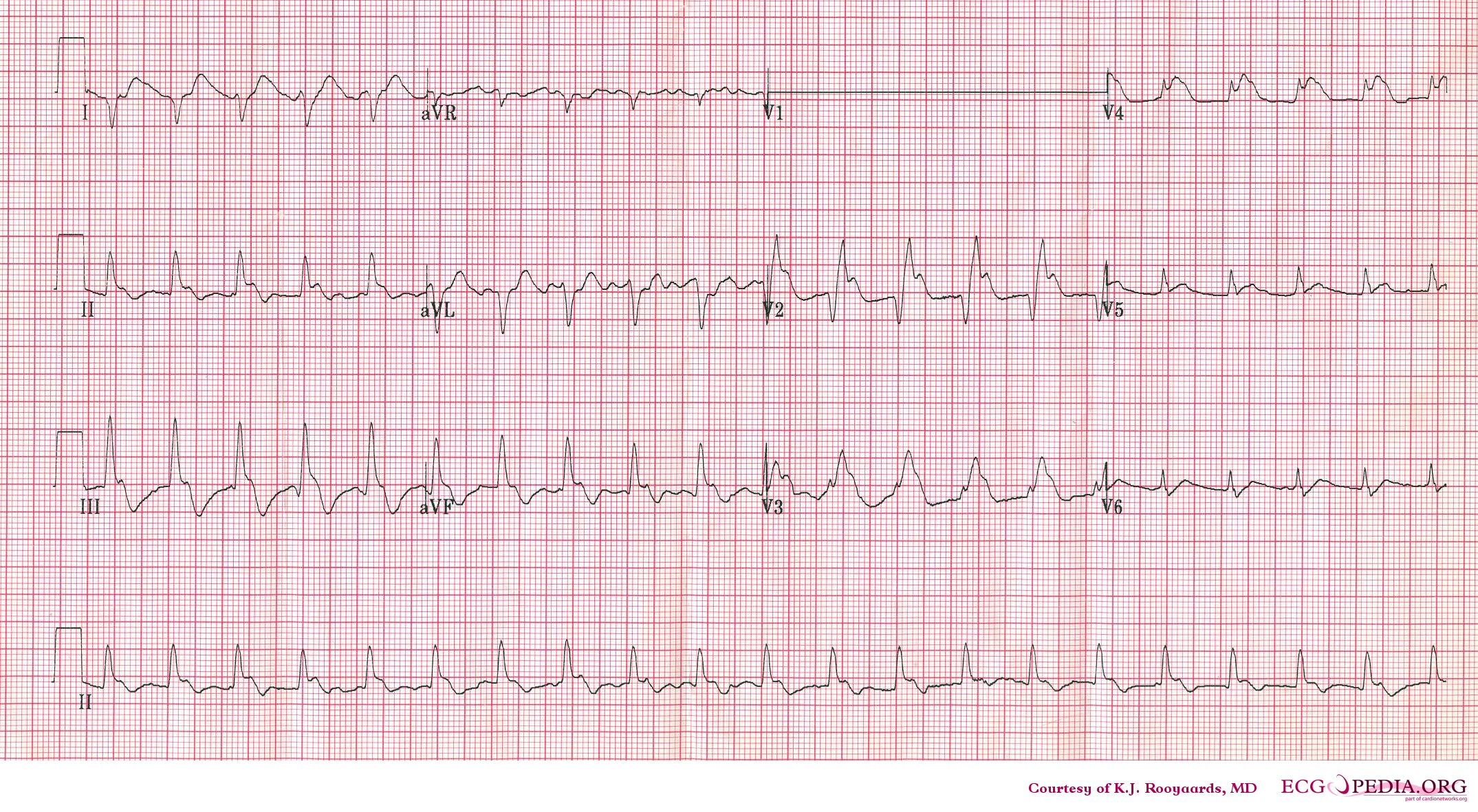
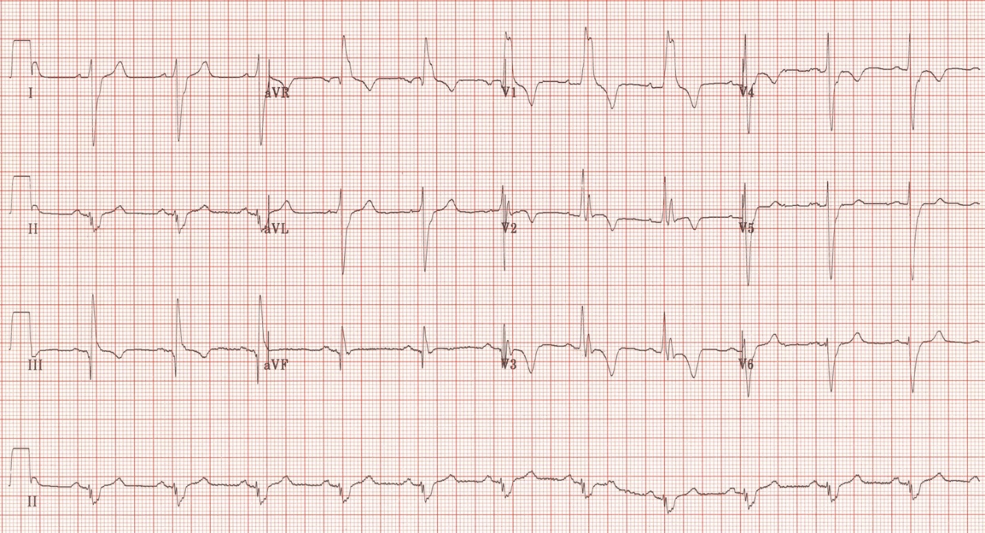
Web electrocardiographic (ecg) abnormalities associated with chronic obstructive pulmonary disease (copd) include right atrial enlargement, right. , md, mhs, johns hopkins university. Web illustration by.

The ECG's of Pulmonary Embolism Resus
These changes can be present on the electrocardiograms of patients. Electrocardiography (ecg) is a useful adjunct to other pulmonary tests because it provides information about.
S1Q3T3 pattern on ECG in pulmonary embolism All About Cardiovascular
Web specific electrocardiographic abnormalities and cardiac arrhythmias are prevalent in chronic obstructive pulmonary disease. The underlying pathophysiology is complex. Web electrocardiographic (ecg) abnormalities associated with.
ECG in Chronic Obstructive Pulmonary Disease • LITFL • ECG Library
Again, this indicates significant right ventricular strain. Web the ecg changes associated with acute pulmonary embolism may be seen in any condition that causes acute.

S1Q3T3 EKG Classic Pattern in Pulmonary Embolism (Example). Pulmonary
Our aim was to separate the effects on ecg by airway obstruction,. Web electrocardiographic (ecg) abnormalities associated with chronic obstructive pulmonary disease (copd) include right.

Pulmonary Embolism (PE) Causes, symptoms, diagnosis, treatment
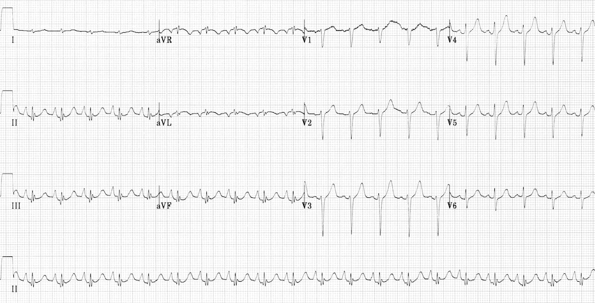
Web ecg abnormalities are common in patients with pulmonary embolism, with the most frequent being sinus tachycardia, right ventricular strain, and the classic s1q3t3 pattern..

Pulmonary embolism and S1Q3 pattern Cardiocases
Again, this indicates significant right ventricular strain. , md, mhs, johns hopkins university. Web specific electrocardiographic abnormalities and cardiac arrhythmias are prevalent in chronic obstructive.
Electrocardiography (Ecg) Is A Useful Adjunct To Other Pulmonary Tests Because It Provides Information About The Right Side Of.
Web sinus tachycardia is the most common ecg finding in pulmonary embolism. Chronic lung disease (cor pulmonale) congenital heart disease (e.g. Web electrocardiography (ecg) in pulmonary disorders. Web a better understanding of the ecg changes in copd may improve interpretation of ecg in these patients and help revealing the dominant pathophysiology of their airway disease.
Associated With Increased Pulmonary Artery Pressures In The Setting Of Acute Or Chronic Right Ventricular Hypertrophy Or Dilatation:
To evaluate the extent and diagnostic values of ecg changes. Ecg changes commonly associated with pulmonary diseases such as copd. This pattern is characterized by a large s wave in lead i, a q wave in lead iii, and an inverted t wave in lead iii. An ecg cannot, by itself, diagnose a pulmonary embolism.
Web Electrocardiography (Ecg) In Pulmonary Disorders.
Electrocardiography (ecg) is a useful adjunct to other pulmonary tests because it. These changes can be present on the electrocardiograms of patients. Web the following ecg signs reflecting ccp were collected: Web copd can cause electrocardiographic changes due to factors including lung hyperinflation.
Web Ecg Abnormalities Are Common In Patients With Pulmonary Embolism, With The Most Frequent Being Sinus Tachycardia, Right Ventricular Strain, And The Classic S1Q3T3 Pattern.
Web the ecg criteria for lvh shown in table 1 have evolved over the years. Web electrocardiographic (ecg) findings may help in clinical decision making regarding this disease entity. Web illustration by maya chastain. The underlying pathophysiology is complex.