Cholestatic Pattern Lfts - Cholestatic pattern of serum enzyme elevations ( r value <2), with alk p levels greater than 3 times uln (>345 u/l) at the time of peak alt or bilirubin elevation. These include the hepatocellular pattern, the cholestatic pattern, and the isolated hyperbilirubinemia pattern. Compared to hepatocellular injury, cholestatic dili is more likely to result in chronicity of insult. Web most of the disease entities can be categorized into hepatocellular or cholestatic patterns, with characteristic traits on liver blood tests. Web although the term liver function tests (lfts) is used commonly, it is imprecise and potentially misleading since many of the tests reflecting the health of the liver are not direct measures of its function. Abnormal liver enzyme levels may signal liver. Upper limit of normal alt. Web an elevation in alp and bilirubin in disproportion to alt and ast would characterize a cholestatic pattern. No reliable autoantibodies have been identified and the diagnosis of psc is usually made on cholestatic liver biochemistry and typical magnetic resonance cholangiopancreatography appearances of intra. The ast:alt ratio may be helpful in determining both the aetiology and the stage of liver disease (see main text).
Liver function tests in primary care bpacnz
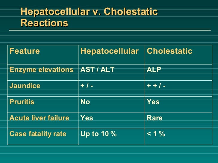
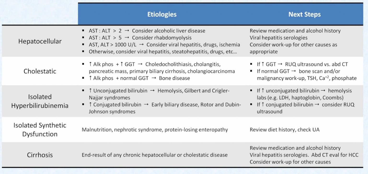
Web there are two main patterns of lft abnormalities that can provide clues about the location or type of liver dysfunction: Compared to hepatocellular injury,.
Liver function tests indication and interpretation The
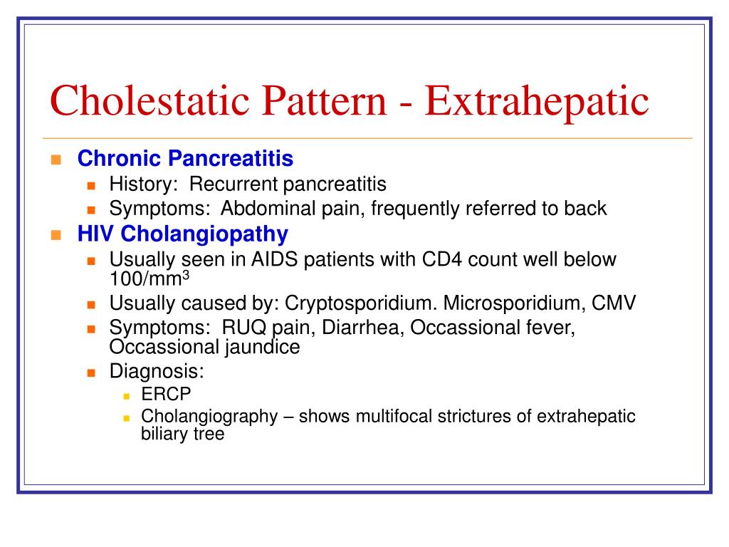
Intrahepatic cholestasis is caused by defects in bile canaliculi, hepatocellular function, or intrahepatic bile ducts. Upper limit of normal alt. A mixed injury pattern is.

PPT Abnormal LFTs PowerPoint Presentation, free download ID139175
The course of drug induced liver injury is considered mixed if features of both hepatocellular (acute hepatitis) and cholestatic injury (cholestatic hepatitis) are present. Cholestatic.

PPT Abnormal LFTs PowerPoint Presentation, free download ID139175
Liver function tests (lfts) are useful blood tests to help identify liver disease, but their interpretation can be challenging. The pattern of alt to alp.

Pin on Infographics
Isolated hyperbilirubinemia is defined as an elevation of bilirubin with normal alkaline phosphatase and ast/alt levels. Web an elevation in alp and bilirubin in disproportion.
PPT Abnormal LFTs PowerPoint Presentation, free download ID139175
A mixed injury pattern is defined as an elevation of alkaline phosphatase and ast/alt levels. Primary biliary cholangitis (pbc) and primary sclerosing cholangitis (psc) are.
Liver Failure Case
Use the first lab values (alt and alp) indicating acute liver injury to calculate the r factor. Intrahepatic cholestasis is caused by defects in bile.

PPT Abnormal LFTs PowerPoint Presentation, free download ID139175
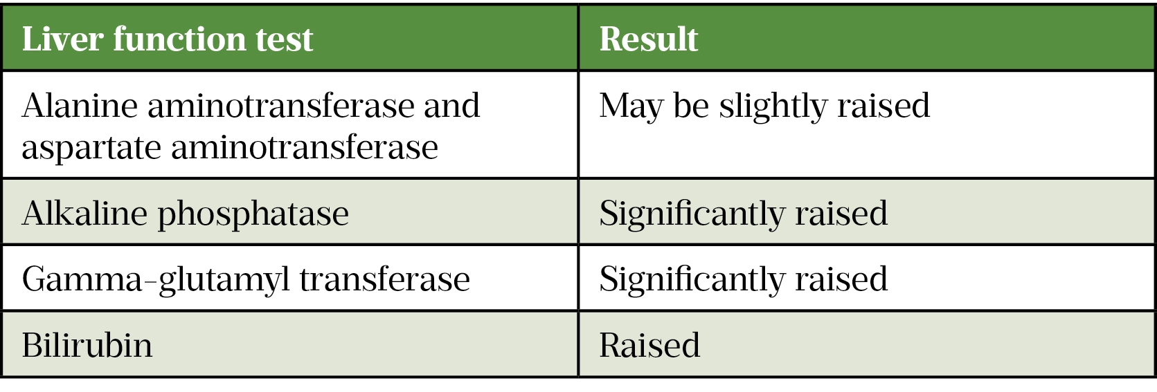
Web last update 26th jan 2021. Web typically, psc causes a chronically abnormal and fluctuant liver function tests (lfts) with a cholestatic pattern (raised alp.
Liver Function Test (LFTs) Normal values, when to order
Web determine the pattern of lft derangement. Web < prev next > mixed hepatitis. Isolated hyperbilirubinemia is defined as an elevation of bilirubin with normal.

LFTs explained Emergency Medicine Kenya Foundation
Isolated hyperbilirubinemia is defined as an elevation of bilirubin with normal alkaline phosphatase and ast/alt levels. Web most of the disease entities can be categorized.
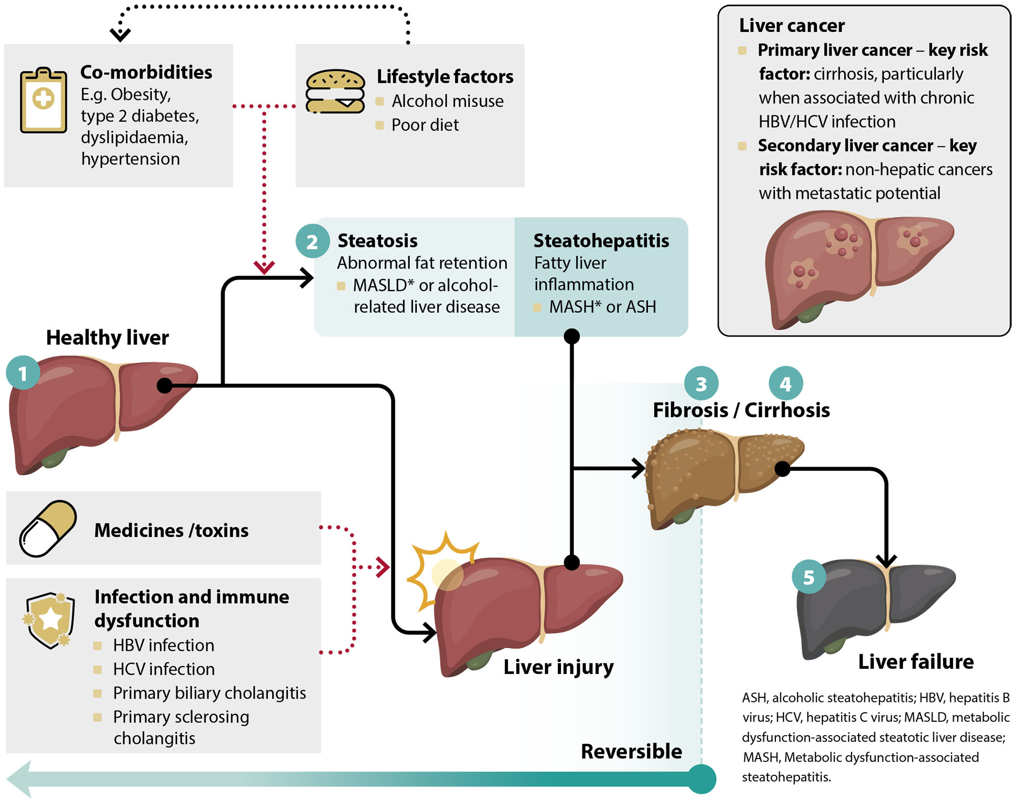
Web Hyperbilirubinemia May Occur As Disease Progresses, And May Lead To Liver Cirrhosis, Liver Failure, Or Even Death.
The pattern of alt to alp rise can indicate whether the pathology is primarily cholestatic or hepatocellular: Use the first lab values (alt and alp) indicating acute liver injury to calculate the r factor. A mixed injury pattern is defined as an elevation of alkaline phosphatase and ast/alt levels. Liver function tests (lfts) are useful blood tests to help identify liver disease, but their interpretation can be challenging.
Web < Prev Next > Mixed Hepatitis.
Web conjugated (direct) bilirubin reflects severe hepatocellular injury or obstruction (cholestatic injury) synthetic liver function is based on proteins made by the liver, including albumin, pt, ptt, platelet count (not discussed here) signs and symptoms. Web an elevation in alp and bilirubin in disproportion to alt and ast would characterize a cholestatic pattern. These include the hepatocellular pattern, the cholestatic pattern, and the isolated hyperbilirubinemia pattern. No reliable autoantibodies have been identified and the diagnosis of psc is usually made on cholestatic liver biochemistry and typical magnetic resonance cholangiopancreatography appearances of intra.
Isolated Hyperbilirubinemia Is Defined As An Elevation Of Bilirubin With Normal Alkaline Phosphatase And Ast/Alt Levels.
1, 2 hepatobiliary diseases with cholestasis from various causes are called cholestatic liver diseases, and cholestasis itself further aggravates liver damage in these patients. Cholestatic liver disease results from insufficient bile synthesis, secretion and/or flow through the biliary tract. Web there are two main patterns of lft abnormalities that can provide clues about the location or type of liver dysfunction: Web using a schematic approach that classifies enzyme alterations as predominantly hepatocellular or predominantly cholestatic, we review abnormal enzymatic activity within the 2 subgroups, the most common causes of enzyme alteration and suggested initial investigations.
The Course Of Drug Induced Liver Injury Is Considered Mixed If Features Of Both Hepatocellular (Acute Hepatitis) And Cholestatic Injury (Cholestatic Hepatitis) Are Present.
Now, let’s focus on the cholestatic pattern. Cholestatic pattern of serum enzyme elevations ( r value <2), with alk p levels greater than 3 times uln (>345 u/l) at the time of peak alt or bilirubin elevation. Web differentiates cholestatic from hepatocellular liver injury, recommended by acg guidelines. How can i recognize a cholestatic pattern?