Bronchial Pattern - Arteries are dorsal and veins are ventral to related bronchi. However, the performance of ai models is contingent upon the datasets used for their training and validation. Web an acb is a supernumerary bronchus from the inner wall of the right main bronchus or intermediate bronchus that progresses toward the pericardium. Vet talks is a project by the ivsa standing committee on veterinary education (scove). Web a bronchial pattern on radiographs indicates a condition that involves the airways. Excessive number of opaque rings and lines, best recognized in the periphery of the lungs where normal. Web a total of 1200 regions of interest (rois) including four specific lung patterns (normal, alveolar, bronchial, and unstructured interstitial) were obtained from 512 thoracic radiographs of 252 dogs and 65 cats. Bronchitis may be either acute or chronic. In a true bronchial pattern that stems from infectious/inflammatory disease, the bronchial walls are thickened because of inflammatory tissue and cells surrounding the airways. An interstitial pattern reflects increased opacity of the pulmonary interstitium.
Interstitial vs Alveolar Lung Patterns wikiRadiography
Arteries are dorsal and veins are ventral to related bronchi. A bronchial pattern is characterized by “doughnuts” and “tramlines”; Bronchitis may be either acute or.

Pulmonary vascular anatomy & anatomical variants. Semantic Scholar
Bacterial > allergic (eosinophilic) cats: Web a bronchial pattern on radiographs indicates a condition that involves the airways. Ai algorithms may accurately classify nsclc. Web.

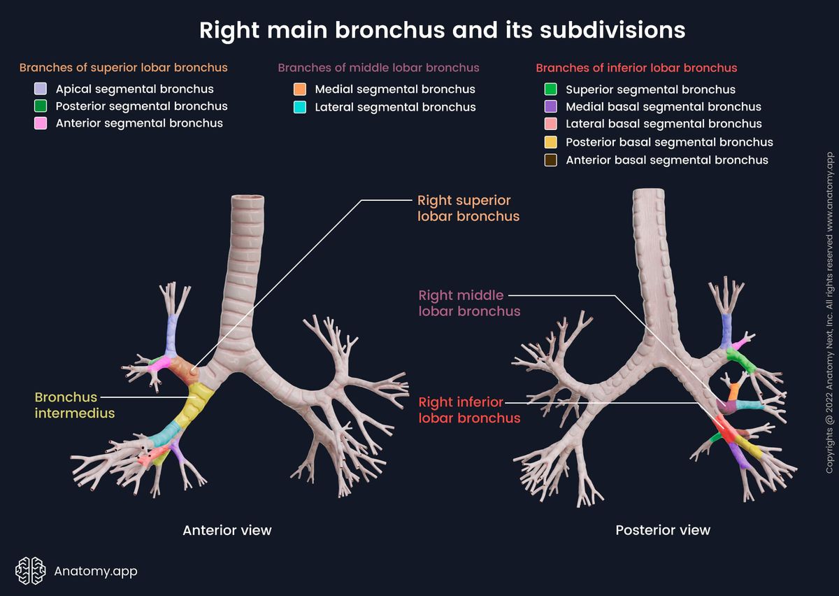
Scheme and nomenclature of the human bronchial tree. Right Main
Bronchitis may be either acute or chronic. In a true bronchial pattern that stems from infectious/inflammatory disease, the bronchial walls are thickened because of inflammatory.
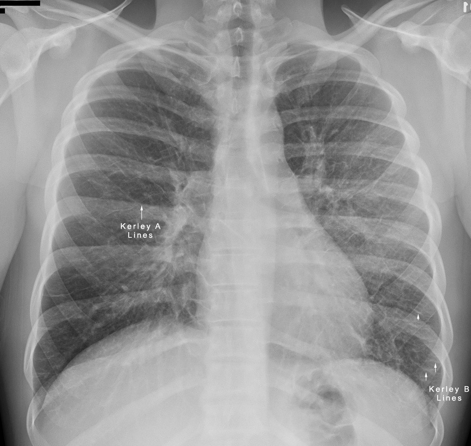
The Radiology Assistant Chest XRay Lung disease
Web bronchitis is an inflammation of the lining of your bronchial tubes, which carry air to and from your lungs. Web every generation, starting from.

Lobar and segmental bronchial anatomy of the Right lung Anatomy
Ai algorithms may accurately classify nsclc. Finally, we describe the spectrum of histologic patterns that are seen in patients with bronchiolar disease who undergo a.
Bronchi Encyclopedia Anatomy.app Learn anatomy 3D models
Vet talks is a project by the ivsa standing committee on veterinary education (scove). Web the three principles of the bronchi nomenclature are as follows:.

Etiologies of pulmonary infections according to CTscan GrepMed
Web an acb is a supernumerary bronchus from the inner wall of the right main bronchus or intermediate bronchus that progresses toward the pericardium. Bronchial.

Lung; cat No. 1. Diffuse, severe bronchointerstitial pattern
The walls are thickened due to a combination of smooth muscle hypertrophy, mucus production, cellular infiltrate, and in come cases (feline asthma), bronchoconstriction. Web the.

Bronchioles Structure
After sixth generation, the passageways are too narrow to be supported by the cartillage, and thus are called bronchioles (small bronchi). This makes them easier.
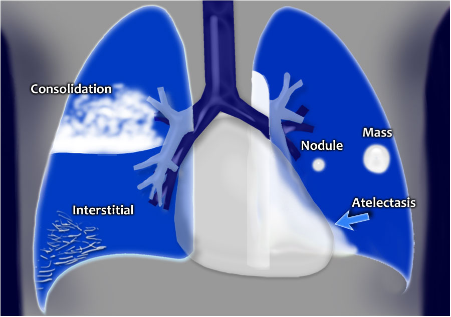
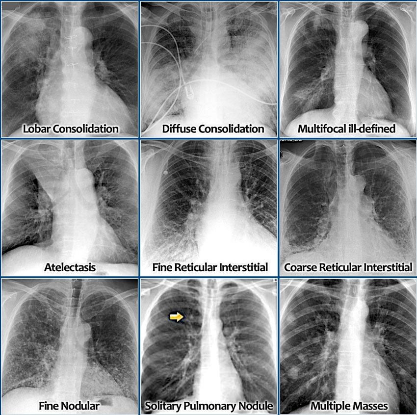
Chest XRay Lung disease FourPattern Approach NCLEX Quiz
Yellow circles) and parallel lines (“tramlines”; It can be a subtle pattern to recognize, so lets look at some of the features. Dorsal (posterior) →.
A Stepwise Approach Allows For Ease And Reliability Of Interpretation ( Figure 2.
It can be a subtle pattern to recognize, so lets look at some of the features. Excessive number of opaque rings and lines, best recognized in the periphery of the lungs where normal. Lateral side → medial side. A bronchial pattern is characterized by “doughnuts” and “tramlines”;
48K Views 7 Years Ago Diagnostic Imaging.
Dorsal (posterior) → ventral (anterior) 3. The bronchial pattern is caused by pathologies (mainly of inflammatory origin) that cause a thickening of the bronchial walls or by pathological infiltration of the peribronchial space. The incidence of t790m mutation at baseline was low in our cohort (2.59%). Web lung cancer is a very aggressive and highly prevalent disease worldwide, with an estimated 2.2 million new cases and 1.8 million deaths in 2020 1.primary lung cancers are divided into two major.
Web The Small Airways (Bronchioles With Inner Diameters <2 Mm), Located At The Transitional Zone Between Larger Conducting Airways And The Pulmonary Acinus, Have Been Overlooked As Major Contributors To Clinical Syndromes Of Respiratory Disease In Cats.
Arteries are dorsal and veins are ventral to related bronchi. Web the tracheobronchial tree is composed of the trachea, the bronchi, and the bronchioles that transport air from the environment to the lungs for gas exchange. Cranial lung lobe vessels are best assessed from the lateral projection; Web the bronchial structure begins at the transverse thoracic plane (also known as the sternal angle at the fourth thoracic vertebra), where the trachea bifurcates into two main bronchi, one for each lung.
Ai Algorithms May Accurately Classify Nsclc.
Radiographic signs of a bronchial pulmonary pattern are: This makes them easier to see, especially in the periphery of the lung (image 2). Once the validity of the spirometry test has been confirmed, the process of interpretation begins. The walls are thickened due to a combination of smooth muscle hypertrophy, mucus production, cellular infiltrate, and in come cases (feline asthma), bronchoconstriction.