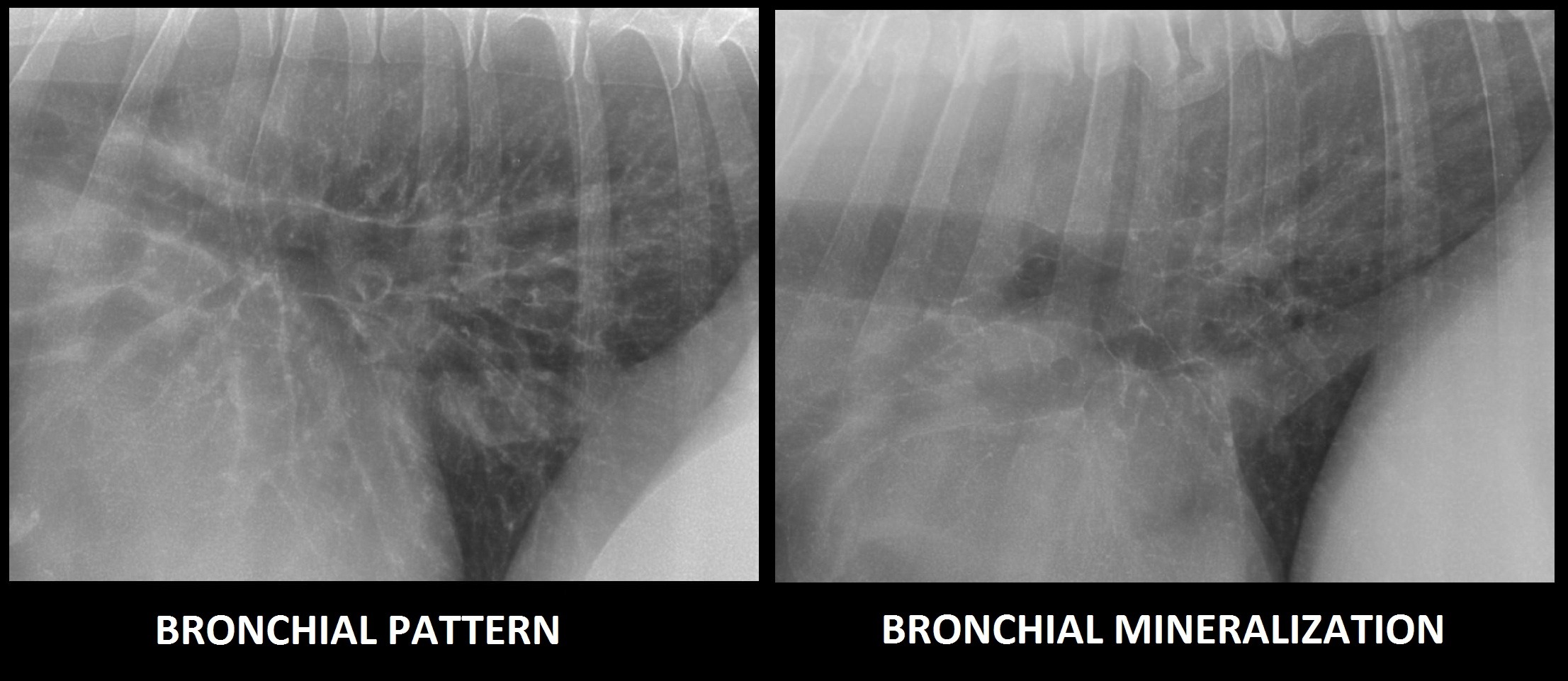
Bronchial Lung Pattern Dog - Interstitial patterns indicate disease or disruption of the interstitium. Web b) bronchial patterns: The thickening of those structures results in enhanced radiographic visualization of the. Web clinically when faced with a mixed pattern, identify the most severe ( i.e. It may also extend into the lungs. Web vet talks is a project by the ivsa standing committee on veterinary education (scove).this vet talk is by dr pete mantis, dvm, dipecvdi, fhea, mrcvs, senior. Web bronchial lung pattern the bronchial pattern is obtained when the bronchial wall is infiltrated by cells or fluid or when the peribronchial space is replaced by cells or fluid. It often occurs in dogs already affected by respiratory disease or a disorder of the lungs or airways. In a true bronchial pattern that stems from infectious/inflammatory disease, the bronchial walls are thickened because of inflammatory tissue and cells surrounding the airways. Bronchial pattern is caused by thickening and increased prominence of the bronchial walls, usually secondary to chronic inflammation.

Radiographic Approach to the Coughing Pet • MSPCAAngell
Normal variants causing increased lung opacity expiration: Web a bronchial pattern is an abnormal lung opacity caused by peribronchial cellular, fluid and fibrotic infiltration, or.
Bronchitis Vs Pneumonia X Ray
Impingement on the main stem bronchi by severe left heart enlargement; Web vet talks is a project by the ivsa standing committee on veterinary education.

(a) Left lateral thoracic radiograph of an eightmonthold dog, showing
It may also extend into the lungs. Web clinically when faced with a mixed pattern, identify the most severe ( i.e. Bronchial pattern is caused.
Image
Impingement on the main stem bronchi by severe left heart enlargement; Radiographic signs of a bronchial pulmonary pattern are: The thickening of those structures results.
Thoracic radiographs from case study 1 demonstrate a multifocal
Web primary lung tumors usually originate from the terminal bronchioles and alveoli; Heart worm disease, large airway disease; Web b) bronchial patterns: Impingement on the.
Thoracic Imaging The Pulmonary Parenchyma • MSPCAAngell
Web b) bronchial patterns: Web tracheobronchitis is a sudden or longterm inflammation of the trachea and bronchial airways; Web clinically when faced with a mixed.

Figure 1 from Topographical distribution and radiographic pattern of
Web b) bronchial patterns: Web tracheobronchitis is a sudden or longterm inflammation of the trachea and bronchial airways; Web clinically when faced with a mixed.

Thoracic radiograph of dog showed mild bronchial pattern (A) and an
Heart worm disease, large airway disease; Web b) bronchial patterns: This makes them easier to see, especially in. A bronchial pattern is important to recognize,.

Topographical distribution and radiographic pattern of lung lesions in
In humans, lung congestion scores are predictive of recurrence of acute congestive heart failure (chf) and are superior to. It may also extend into the.

Clinician's Brief Canadian Edition September 2020 Digital Edition
Web vet talks is a project by the ivsa standing committee on veterinary education (scove).this vet talk is by dr pete mantis, dvm, dipecvdi, fhea,.
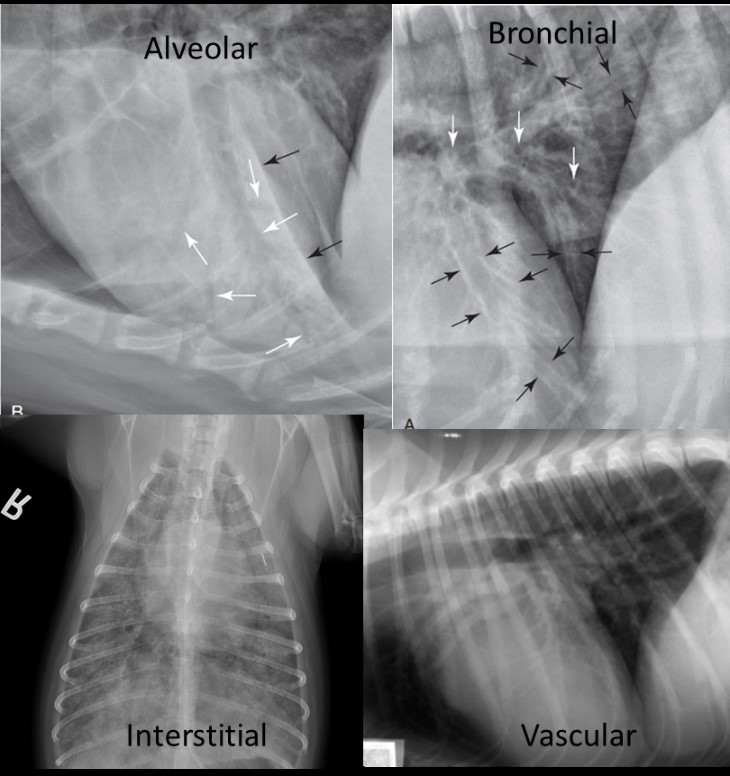
Web In Addition To Examination Of Extrathoracic Structures, The Pleural And Mediastinal Space, And Heart Size And Shape, Careful Attention Should Be Given To Pulmonary Pattern (E.g., Alveolar, Interstitial, Bronchial, Or Vascular), Distribution (E.g., Affected Lobes,.
Web bronchial lung pattern the bronchial pattern is obtained when the bronchial wall is infiltrated by cells or fluid or when the peribronchial space is replaced by cells or fluid. Web four different patterns of increased lung opacity have been described in order to classify different disease processes: This pattern comes closest to helping shed light on what disease the pet is suffering from. Characteristic findings include an increased opacity in the lungs that partially obscures blood vessel margins, which may be due to the presence of.
A Bronchial Pattern Is Important To Recognize, Because, While It May Be A Normal Variant In An Aged Patient, It May Also Be Due To A.
This makes them easier to see, especially in. Web b) bronchial patterns: In humans, lung congestion scores are predictive of recurrence of acute congestive heart failure (chf) and are superior to. Web a bronchial pattern is diffuse thickening of the airway walls giving the appearance of thick lines and rings throughout the lungs.
The Walls Are Thickened Due To A Combination Of Smooth Muscle Hypertrophy, Mucus Production,.
Radiographic signs of a bronchial pulmonary pattern are: Normal variants causing increased lung opacity expiration: Web a bronchial pattern is an abnormal lung opacity caused by peribronchial cellular, fluid and fibrotic infiltration, or bronchial mucosal and submucosal thickening (chronic bronchitis). Web clinically when faced with a mixed pattern, identify the most severe ( i.e.
The Thickening Of Those Structures Results In Enhanced Radiographic Visualization Of The.
Impingement on the main stem bronchi by severe left heart enlargement; Interstitial patterns indicate disease or disruption of the interstitium. Web vet talks is a project by the ivsa standing committee on veterinary education (scove).this vet talk is by dr pete mantis, dvm, dipecvdi, fhea, mrcvs, senior. They occasionally develop as a second coincidental tumor, which can make differentiation between primary and metastatic disease difficult.