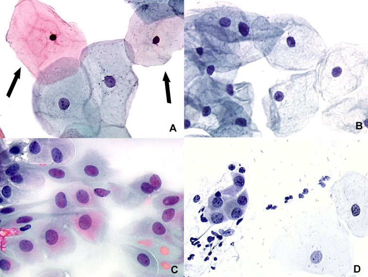
Atrophic Pattern Predominantly Parabasal Cells - Naked nuclei (small cells) may be seen. Web degenerating parabasal cells, pseudoparakeratosis, and necrotic background are associated with atrophic vaginitis ( p =.001) on papanicolaou. Web a pap smear is used to screen for cervical cancer. Vaginal atrophy in menopause shows increased parabasal cells on cytology. 3 c, d), further complicating the interpretation. External genitalia should be examined for. The health care professional first places a speculum inside the vagina. Loss of fragile cytoplasm of the thin atrophic and relatively dry epithelium leads to plenty bare nuclei throughout the smear. How is a pap test done? Web the occurrence of vaginal atrophy during menopause is associated with declining estrogen levels that cause structural and functional changes in vaginal tissue, including atrophy of vaginal.
Pathology Outlines Parabasal cells
Web the occurrence of vaginal atrophy during menopause is associated with declining estrogen levels that cause structural and functional changes in vaginal tissue, including atrophy.

Parabasal cells in pap smear with postpartum Ad , ad, cells
Web atrophic pattern histologic findings demonstrate decreased superficial squamous cells, increased parabasal cells, decreased lactobacilli. When to see a doctor. 3 c, d), further complicating.
Cytopathology of the uterine cervix digital atlas
The pap smear is usually done in conjunction with a pelvic exam. Naked nuclei (small cells) may be seen. This results in itching, burning and.

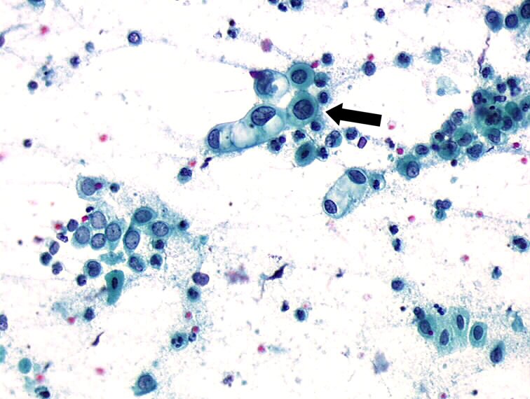
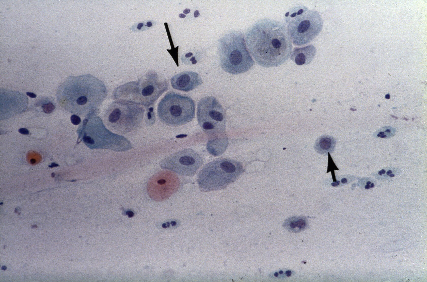
− Atrophy associated with inflammation A) parabasal squamous epithelial
How is a pap test done? A doctor has provided 1 answer. Web vaginal atrophy is a condition where the lining of your vagina gets.
Histopathology and cytopathology of the uterine cervix digital atlas
In women older than age 30, the pap test may be combined with a test for human papillomavirus (hpv) — a common sexually transmitted infection.
Pap Smear, Parabasal Cells Photograph by Science Source
Web pap test is an important screening test for cervical cancer. Loss of fragile cytoplasm of the thin atrophic and relatively dry epithelium leads to.

− Atrophy associated with inflammation A) parabasal squamous epithelial
Atrophic pattern predominantly parabasal cells. The health care professional first places a speculum inside the vagina. Web furthermore, recognizing the parabasal cells in the menopausal.
Parabasal cells Collection
However, there are normal to low numbers of neutrophils. Pseudokeratinized cells (pink to orangophilic cytoplasm) are due to degeneration. How is a pap test done?.

Cytopathology of the uterine cervix digital atlas
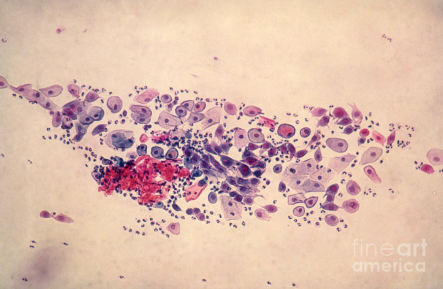
Web the smear pattern of an atrophic smear with marked inflammation comprises sheets of and dissociated parabasal cells. This results in itching, burning and pain.

− Atrophy associated with inflammation A) parabasal squamous epithelial
External genitalia should be examined for. While evaluating the cytomorphologic features of a pap smear from the cervix or vagina, it is important to know.
Web Atrophic Epithelium Appears Pale, Smooth And Shiny.
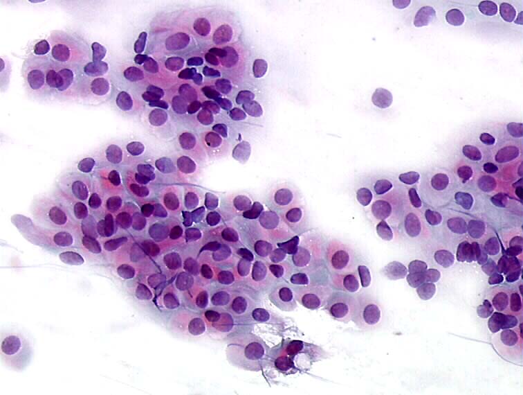
External genitalia should be examined for. Web furthermore, recognizing the parabasal cells in the menopausal smears, either singly or as syncytial aggregates, is important to avoid overdiagnosis of squamous intraepithelial lesions. This may be accompanied by abundant neutrophils. Web atrophic pattern histologic findings demonstrate decreased superficial squamous cells, increased parabasal cells, decreased lactobacilli.
Web Vaginal Atrophy Is A Condition Where The Lining Of Your Vagina Gets Drier And Thinner.
The condition also includes urinary tract problems such as urinary tract infections (utis) and urinary incontinence. The health care professional first places a speculum inside the vagina. However, there are normal to low numbers of neutrophils. Pseudokeratinized cells (pink to orangophilic cytoplasm) are due to degeneration.
Vaginal Atrophy (Atrophic Vaginitis) Is Thinning, Drying And Inflammation Of The Vaginal Walls That May Occur When Your Body Has Less Estrogen.
In atrophic smears, parabasal and some basal cells are the characteristic cell types. Loss of fragile cytoplasm of the thin atrophic and relatively dry epithelium leads to plenty bare nuclei throughout the smear. Atrophic pattern predominantly parabasal cells. Vaginal atrophy in menopause shows increased parabasal cells on cytology.
When To See A Doctor.
This results in itching, burning and pain during sex, among other symptoms. A shift in maturation index in the absence of significant inflammation is more accurately termed atrophic pattern. Web pap test is an important screening test for cervical cancer. 3 c, d), further complicating the interpretation.