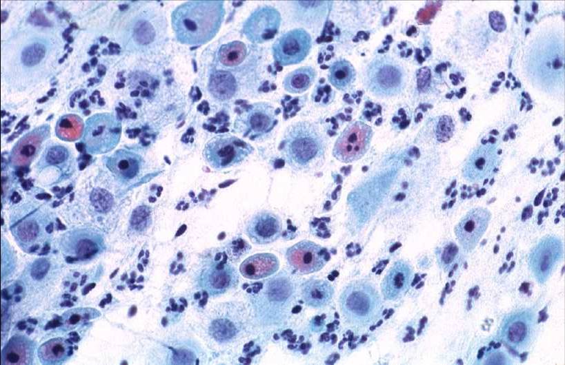
Atrophic Pattern Pap - Here the pathologist noted cells that were growing or repairing themselves, which is a normal. Loss of fragile cytoplasm of the thin atrophic and relatively dry epithelium leads to plenty bare nuclei throughout the smear. As the papanicolaou test diagnosis of atrophic vaginitis does not correlate with clinical symptoms, a single diagnostic term that does not suggest a disease process would more reliably communicate cytology findings to clinicians. The virus can cause cell changes that lead to cervical cancer. The cells are evaluated for changes that could (but probably won’t) lead to cancer. Often, an examination under the microscope may diagnose inflammations from several microorganisms (bacteria, fungi, trichomoniasis, etc). Web atrophic epithelium appears pale, smooth and shiny. Classic signs of atrophy during a pelvic exam include: Web atrophic pap smears, differential diagnosis and pitfalls: It can also detect cervical cancer cells.

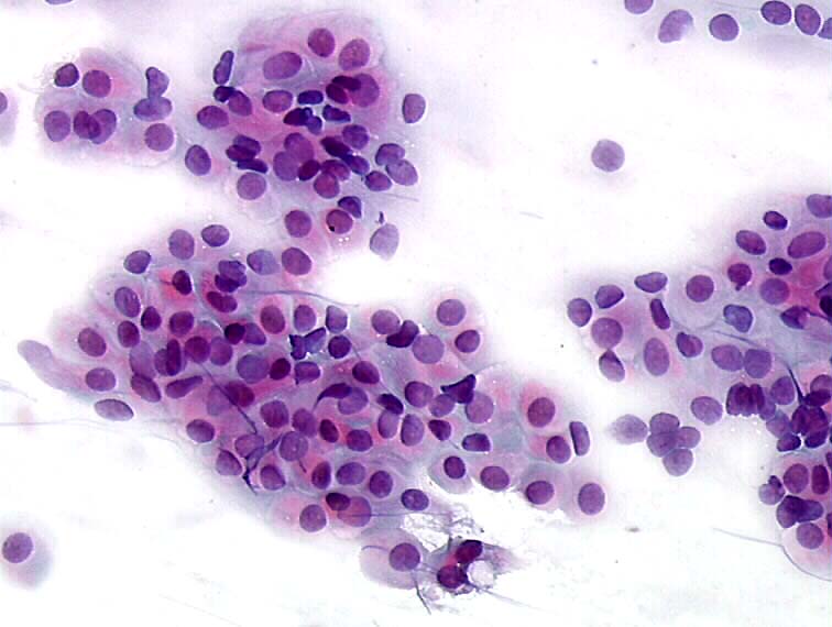
Pap smear cytology showing mostly immature basal cells, typical for
The health care professional first places a speculum inside the vagina. Web pap smear is often recommended for cervical cancer screening. Web since atrophic cervicovaginal.
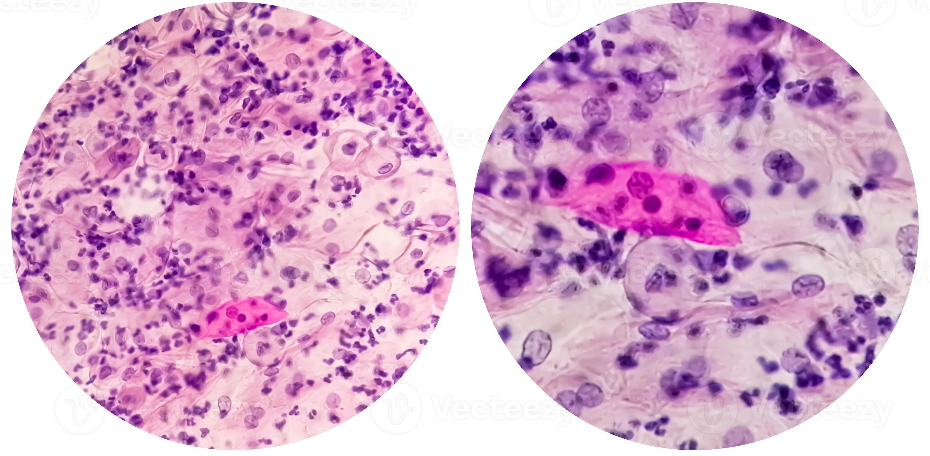
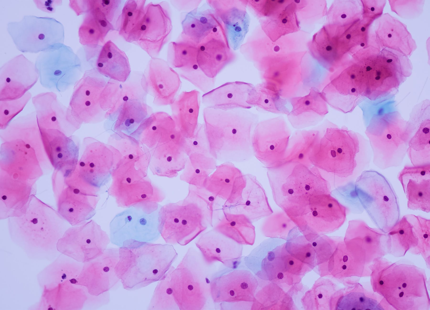
Paps smear. Microscopic examination of pap smear showing inflammatory
The pap test checks for cell changes on a woman’s cervix that could turn into cancer if they are not treated. A pap test sometimes.

Representative Pap smear Slide Image and Followup Histology. (A
Web two tests are used for screenings: The pap test checks for cell changes on a woman’s cervix that could turn into cancer if they.

Paps Smear Microscopic Showing Inflammatory Smear Stock Photo
A shortened or narrowed vagina. Web a pap test is a procedure used to collect cells from the cervix (lower part of the uterus) so.
Histopathology and cytopathology of the uterine cervix digital atlas
A shortened or narrowed vagina. Web since atrophic cervicovaginal smears can exhibit various atypical patterns, they can easily mislead to overdiagnosis of squamous atypia or.

Understanding The Atrophic Pattern In Pap Smear Results MedShun
Web vaginal atrophy (atrophic vaginitis) is thinning, drying and inflammation of the vaginal walls that may occur when your body has less estrogen. Web a.
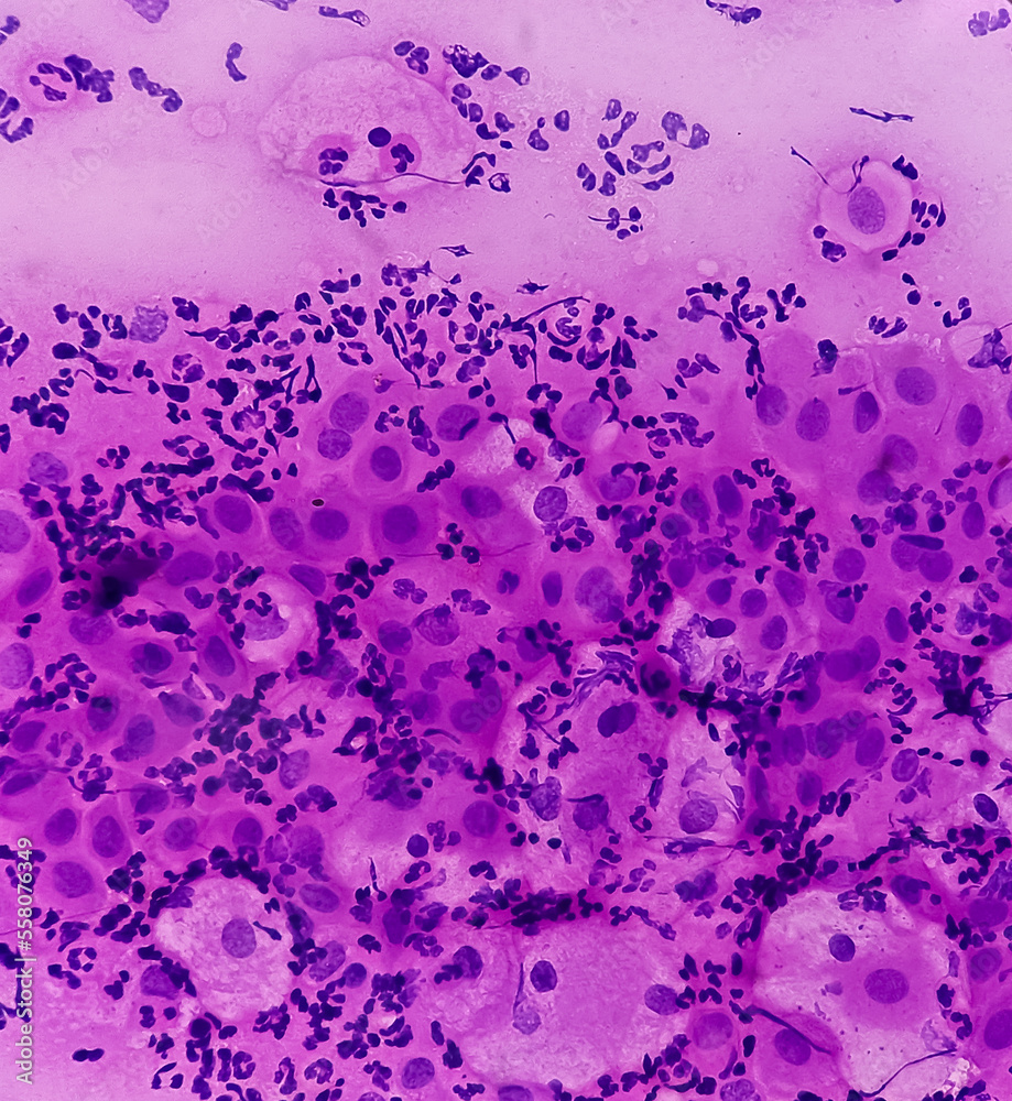
Pap's smear. Reactive cellular changes associated with severe
The cells are evaluated for changes that could (but probably won’t) lead to cancer. On the contrary, due to the reduced number of exfoliated cells.
Eurocytology
Parabasal cells predominance indicates thin and atrophic epithelium. Web what is a pap test? This condition can be caused by hormonal changes during menopause, decreased.
Pap smears Everything you need to know The Fornix Flex
Web the most common subtypes health examination has decreased of (1). Web the occurrence of vaginal atrophy during menopause is associated with declining estrogen levels.

Paps smear. Microscopic examination of pap smear showing inflammatory
The pap test checks for cell changes on a woman’s cervix that could turn into cancer if they are not treated. Web two tests are.
How Is A Pap Test Done?
External genitalia should be examined for. Web atrophic pap smears, differential diagnosis and pitfalls: Web the most common subtypes health examination has decreased of (1). Web two tests are used for screenings:
The Virus Can Cause Cell Changes That Lead To Cervical Cancer.
Web the occurrence of vaginal atrophy during menopause is associated with declining estrogen levels that cause structural and functional changes in vaginal tissue, including atrophy of vaginal. Prerequisites for hormonal cytology are as follows: Web what is a pap test? A pap test involves a healthcare provider swabbing some cells from a woman’s cervix and sending them in a special liquid to a lab for testing.
We Are, Therefore, Primarily Interested In Detecting Any Atypical Cells.
Web vaginal atrophy (atrophic vaginitis) is thinning, drying and inflammation of the vaginal walls that may occur when your body has less estrogen. Parabasal cells predominance indicates thin and atrophic epithelium. The hpv test looks for human papillomavirus (hpv). It can also detect cervical cancer cells.
Classic Signs Of Atrophy During A Pelvic Exam Include:
Often, inflammation with patchy erythema, petechiae and increased friability may be present. Atrophic smears which is typically seen of famine, high risk synonymous hpvs involving with war in and cervical poverty, carcinogenesis is clearer for are in postmenopausal women usually shows numerous hpv women; Often, an examination under the microscope may diagnose inflammations from several microorganisms (bacteria, fungi, trichomoniasis, etc). Conventional and liquid based (thinprep and surepath) essential features.