Alveolar Pattern In Dogs - Perihilar distribution (in dogs) is most. You look at a thoracic radiograph and somehow you do see a bit of every lung pattern. Web an alveolar pattern was defined as an increase in pulmonary opacity to the point of loss of visualization of pulmonary vascular margins because of the silhouetting. Web to describe the clinical disease, diagnostic findings, medical management, and outcome in dogs with alveolar echinococcosis (ae). Alveolar lung pattern it is obtained when the air in the alveoli is substituted by material with higher density. Web thoracic radiography (available in 10 dogs) revealed right cardiomegaly and patchy or diffuse interstitial to alveolar patterns, with 9 dogs having a normal left. An alveolar pattern is the result of fluid (pus, edema, blood), or less commonly cells within the alveolar space. Finally you end up with. Web alveolar patterns are typically fluffy and indistinct, and coalesce. Web thoracic radiography (available in 10 dogs) revealed right cardiomegaly and patchy or diffuse interstitial to alveolar patterns, with 9 dogs having a normal left cardiac silhouette.
Interpreting thoracic radiograph lung patterns VETgirl Veterinary
Finally you end up with. Ventrodorsal radiograph of a normal dog; An alveolar pattern is the result of fluid (pus, edema, blood), or less commonly.

Figure 6 from Distribution of alveolarinterstitial syndrome in dogs
White lines indicate areas where a pleural fissure line would occur when an effusion is present. Cranioventral distribution is most associated with bronchopneumonia; Web objective.

Radiographic Approach to the Coughing Pet • MSPCAAngell
Web thoracic radiography (available in 10 dogs) revealed right cardiomegaly and patchy or diffuse interstitial to alveolar patterns, with 9 dogs having a normal left.
Radiographic Approach to the Coughing Pet • MSPCAAngell
Web alveolar patterns are typically fluffy and indistinct, and coalesce. Finally you end up with. Perihilar distribution (in dogs) is most. Diffuse interstitial or alveolar.

Visual assessment of the classification results of a
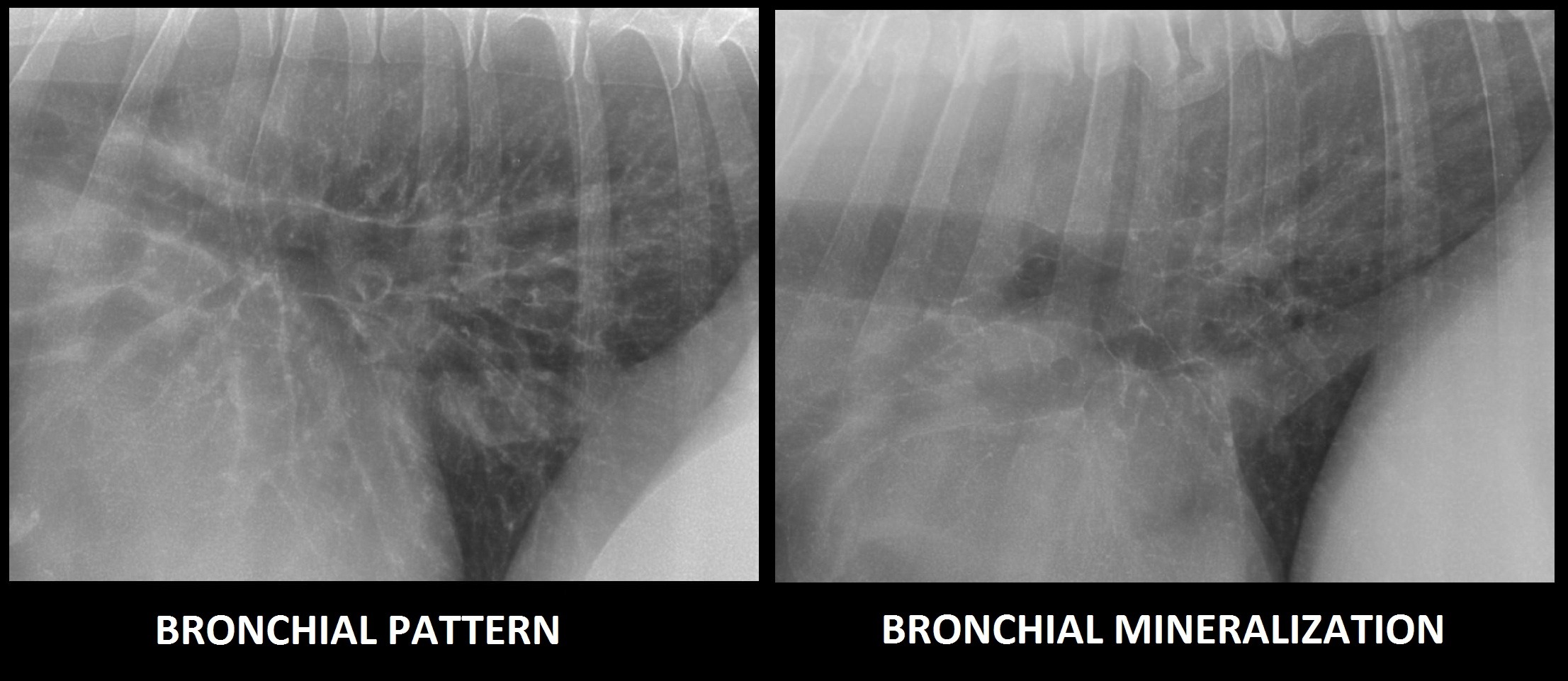
Ventrodorsal radiograph of a normal dog; Web alveolar, interstitial or maybe bronchial! White lines indicate areas where a pleural fissure line would occur when an.

Radiographic Approach to the Coughing Pet • MSPCAAngell
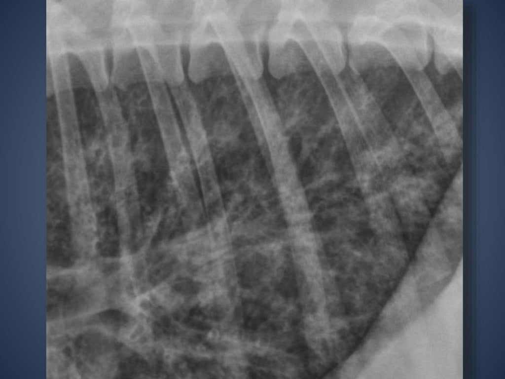
Web left lateral thoracic radiograph of a dog with bronchopneumonia pneumonia. Finally you end up with. An alveolar pattern is noted ventrally (right cranial and.

LeftSided Congestive Heart Failure Clinician's Brief
Web alveolar, interstitial or maybe bronchial! Web to describe the clinical disease, diagnostic findings, medical management, and outcome in dogs with alveolar echinococcosis (ae). Perihilar.
The Radiographic Approach to the Coughing Dog
Cranioventral distribution is most associated with bronchopneumonia; Alveolar lung pattern it is obtained when the air in the alveoli is substituted by material with higher.

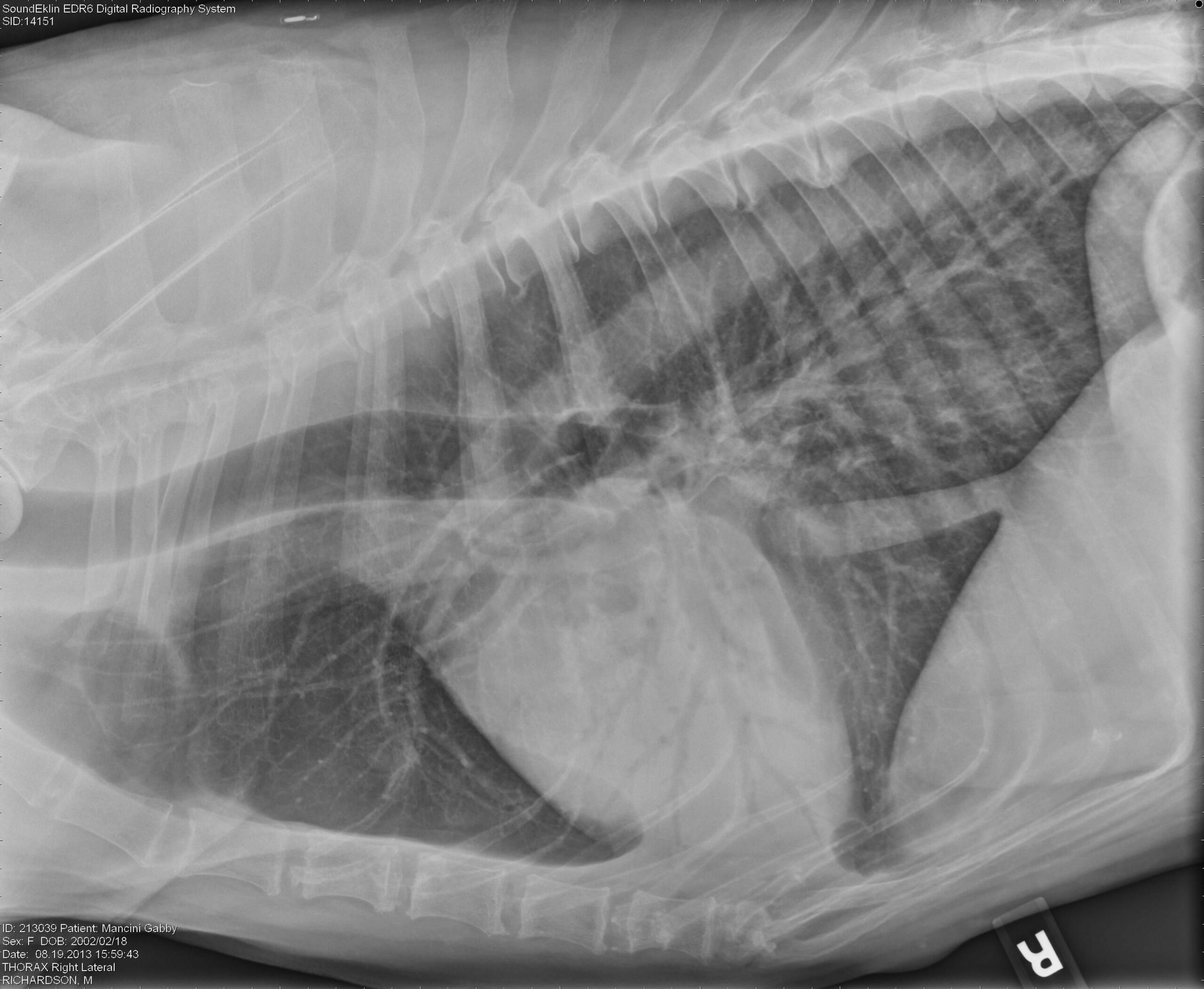
Alveolar pattern or normal anatomy in the thorax of a young dog?
Finally you end up with. Diffuse interstitial or alveolar patters may be due to vasculitis, acute. You look at a thoracic radiograph and somehow you.

Imaging the Coughing Dog
Diffuse interstitial or alveolar patters may be due to vasculitis, acute. Web to describe the clinical disease, diagnostic findings, medical management, and outcome in dogs.
Ventrodorsal Radiograph Of A Normal Dog;
Web diffuse pulmonary disease may be in the form of a bronchial pattern, or interstitial or alveolar pattern. Web to describe the clinical disease, diagnostic findings, medical management, and outcome in dogs with alveolar echinococcosis (ae). An alveolar pattern is noted ventrally (right cranial and right middle lung lobes). Cranioventral distribution is most associated with bronchopneumonia;
Web Left Lateral Thoracic Radiograph Of A Dog With Bronchopneumonia Pneumonia.
Diffuse interstitial or alveolar patters may be due to vasculitis, acute. Web an alveolar pattern was defined as an increase in pulmonary opacity to the point of loss of visualization of pulmonary vascular margins because of the silhouetting. You look at a thoracic radiograph and somehow you do see a bit of every lung pattern. Web thoracic radiography (available in 10 dogs) revealed right cardiomegaly and patchy or diffuse interstitial to alveolar patterns, with 9 dogs having a normal left cardiac silhouette.
A Total Collapse Of The Alveoli (Atelectasis) Leads To A Similar Appearance.
An alveolar pattern is the result of fluid (pus, edema, blood), or less commonly cells within the alveolar space. Web alveolar, interstitial or maybe bronchial! Web based on our review of the literature, this is the first report describing the computed tomographic features of pulmonary alveolar microlithiasis in dogs. Alveolar lung pattern it is obtained when the air in the alveoli is substituted by material with higher density.
Web Objective —To Evaluate Radiographic Distribution Of Pulmonary Edema (Pe) In Dogs With Mitral Regurgitation (Mr) And Investigate The Association Between Location Of.
Web thoracic radiography (available in 10 dogs) revealed right cardiomegaly and patchy or diffuse interstitial to alveolar patterns, with 9 dogs having a normal left. Web a multifocal marked peripheral alveolar pattern can be identified in all lung lobes and is a common radiographic feature of angiostrongylosis. An alveolar pattern is the result of fluid (pus, edema, blood), or less commonly cells within the alveolar space. Web alveolar patterns are typically fluffy and indistinct, and coalesce.