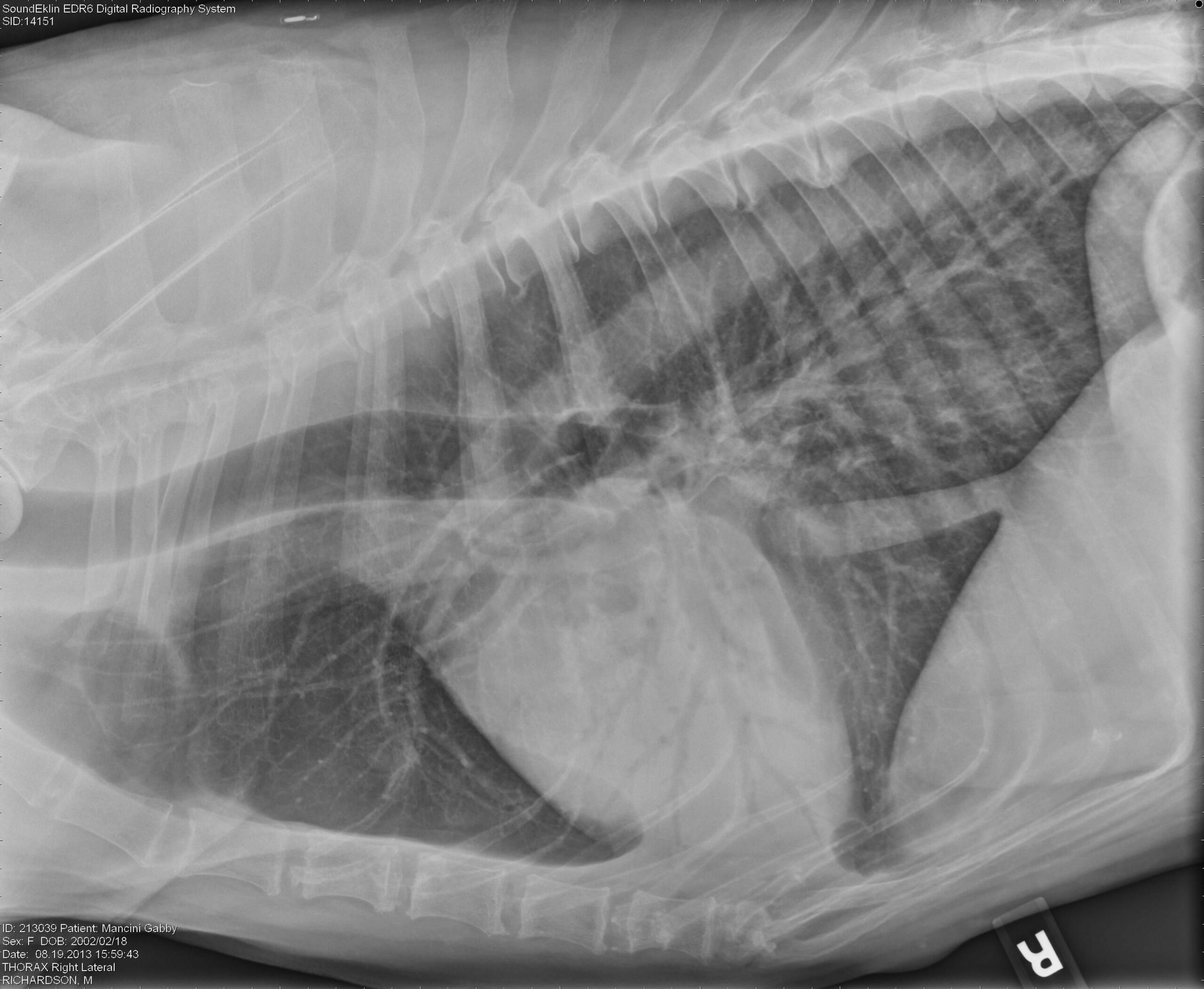
Alveolar Pattern Dog - You look at a thoracic radiograph and somehow you do see a bit of every lung pattern. Multiple air bronchograms are observed in the left hemithorax on. Web an alveolar lung pattern is an opaque lung that completely obscures the margins of the pulmonary blood vessels. The alveolar pattern is the dominant pattern, and will obscure other patterns by silhouette effect. Web learn how to recognize and differentiate common lung patterns and distributions of pulmonary diseases in dogs and cats using thoracic radiographs. Web radiographic evidence of bacterial pneumonia can appear as a focal, multifocal, or diffuse alveolar pattern, although early in the disease process infiltrates. To evaluate the radiographic lung pattern and topographical distribution in canine eosinophilic. The only distinction these patterns make with. Web alveolar, interstitial or maybe bronchial! This pattern results in more loss of airspace than any other pattern.

Visual assessment of the classification results of a
Web radiographic findings varied, but included abnormal unstructured interstitial (one) and unstructured interstitial and alveolar (five) pulmonary patterns, which were. Web certain generalizations have been.
Radiographic Approach to the Coughing Pet • MSPCAAngell
Web radiographic findings used as non mutually exclusive labels to train the cnns were: Unremarkable, cardiomegaly, alveolar pattern, bronchial pattern,. Web alveolar, interstitial or maybe.

Figure 1 from Topographical distribution and radiographic pattern of
Web radiographic evidence of bacterial pneumonia can appear as a focal, multifocal, or diffuse alveolar pattern, although early in the disease process infiltrates. Tracheal disease.

Alveolar pattern or normal anatomy in the thorax of a young dog?
Web thoracic radiography (available in 10 dogs) revealed right cardiomegaly and patchy or diffuse interstitial to alveolar patterns, with 9 dogs having a normal left..

Imaging the Coughing Dog
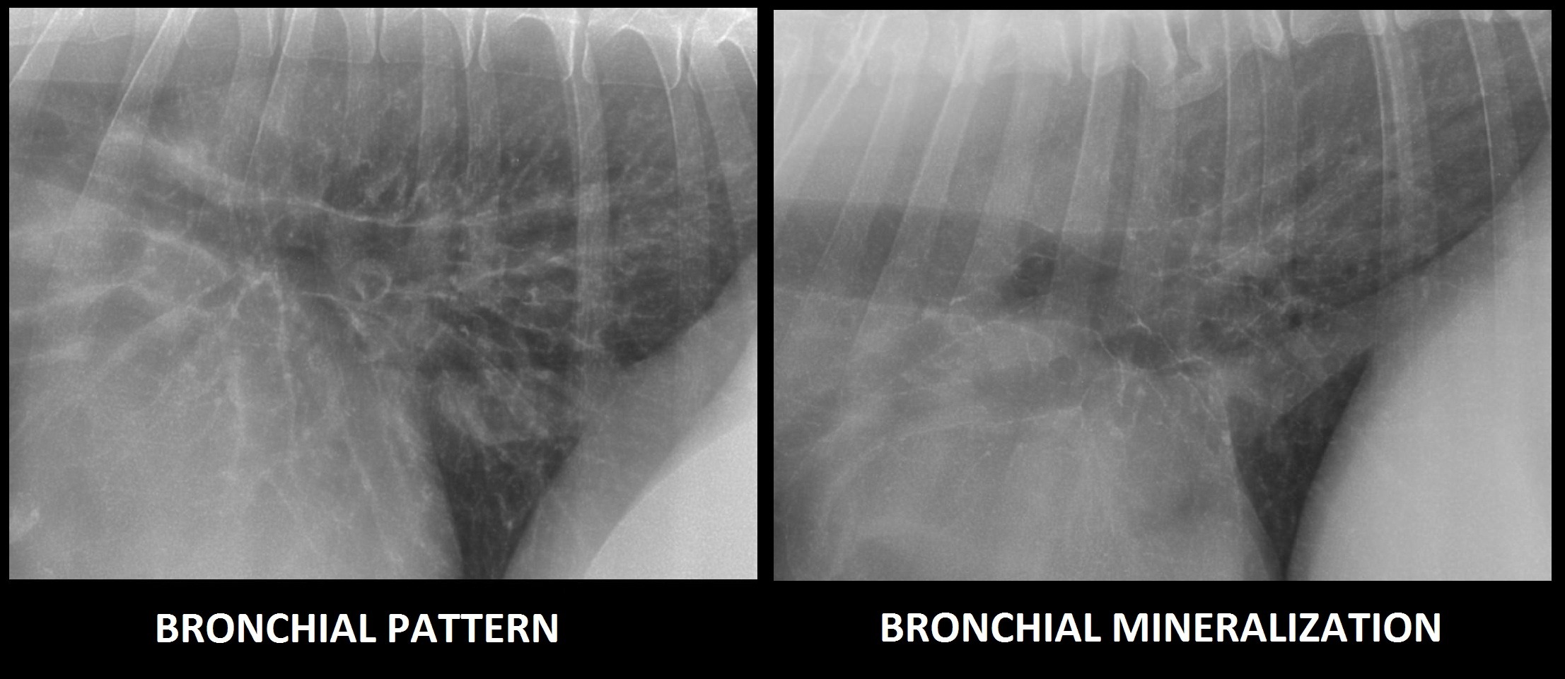
Web an alveolar pattern is the result of fluid (pus, edema, blood), or less commonly cells within the alveolar space. This pattern results in more.
Interpreting thoracic radiograph lung patterns VETgirl Veterinary
To evaluate the radiographic lung pattern and topographical distribution in canine eosinophilic. You look at a thoracic radiograph and somehow you do see a bit.

Radiographic Approach to the Coughing Pet • MSPCAAngell
Tracheal disease may cause dry, honking, resonant cough (dogs) and dyspnea or strider (cats);. The alveolar pattern is the dominant pattern, and will obscure other.

Imaging the Coughing Dog
The only distinction these patterns make with. Unremarkable, cardiomegaly, alveolar pattern, bronchial pattern,. To evaluate the radiographic lung pattern and topographical distribution in canine eosinophilic..

Radiographic Approach to the Coughing Pet • MSPCAAngell
Tracheal disease may cause dry, honking, resonant cough (dogs) and dyspnea or strider (cats);. Web radiographic findings used as non mutually exclusive labels to train.

Figure 6 from Distribution of alveolarinterstitial syndrome in dogs
Unremarkable, cardiomegaly, alveolar pattern, bronchial pattern,. Web an alveolar pattern is the result of fluid (pus, edema, blood), or less commonly cells within the alveolar.
Finally You End Up With.
Web an alveolar pattern in the entire left hemithorax and in the hilar and midzone regions of the right caudal lung lobe. Web certain generalizations have been made about the character of the cough: Multiple air bronchograms are observed in the left hemithorax on. Web radiographic findings used as non mutually exclusive labels to train the cnns were:
Web Important Points Regarding The Alveolar Pattern:
Web radiographic evidence of bacterial pneumonia can appear as a focal, multifocal, or diffuse alveolar pattern, although early in the disease process infiltrates. Web a multifocal marked peripheral alveolar pattern can be identified in all lung lobes and is a common radiographic feature of angiostrongylosis. Web an alveolar pattern is the result of fluid (pus, edema, blood), or less commonly cells within the alveolar space. This pattern results in more loss of airspace than any other pattern.
Unremarkable, Cardiomegaly, Alveolar Pattern, Bronchial Pattern,.
The only distinction these patterns make with. Web an alveolar lung pattern is an opaque lung that completely obscures the margins of the pulmonary blood vessels. Web learn how to recognize and differentiate common lung patterns and distributions of pulmonary diseases in dogs and cats using thoracic radiographs. Web pulmonary alveolar proteinosis, described in dogs and a cat, is a rare disorder resulting from flooding of the alveoli with surfactant.
To Evaluate The Radiographic Lung Pattern And Topographical Distribution In Canine Eosinophilic.
A total collapse of the alveoli (atelectasis) leads to a similar. You look at a thoracic radiograph and somehow you do see a bit of every lung pattern. Tracheal disease may cause dry, honking, resonant cough (dogs) and dyspnea or strider (cats);. Web radiographic findings varied, but included abnormal unstructured interstitial (one) and unstructured interstitial and alveolar (five) pulmonary patterns, which were.