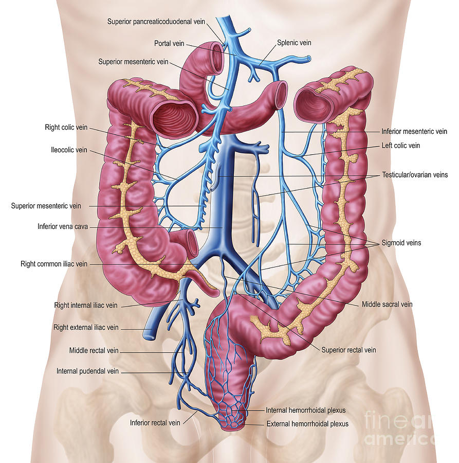
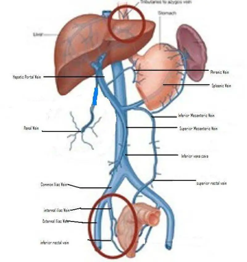
Abdominal Venous Pattern - Checking the abdominal and pelvic venous systems. The portal system transports venous blood to the liver for processing, whilst the systemic venous system returns blood to the right atrium of the heart. Web the recognition of changes in abdominal blood flow allows accurate diagnosis of arterial and venous abnormalities, including stenosis, occlusion, and thrombosis. When liver disease is severe enough to cause cirrhosis, the increase in portal hypertension can lead to backup. Nearly 80% of hepatic inflow comes from the portal vein. The key difference between these two systems is the liver. The portal venous system is composed of the veins that drain the abdominal viscera, spleen, pancreas, and gallbladder. The posterolateral abdominal wall is primarily drained by the tenth and eleventh posterior intercostal, subcostal, and lumbar veins. The inferior vena cava forms at the level of the fifth lumbar vertebra by the joining of left with the right common iliac veins. The world's most advanced 3d anatomy platform.

Veins of Posterior Abdominal Wall Anatomy pediagenosis
The venous drainage of these organs is unlike that of the rest of the body. Flow to the umbilicus (rare, in portal vein thrombosis). Web.
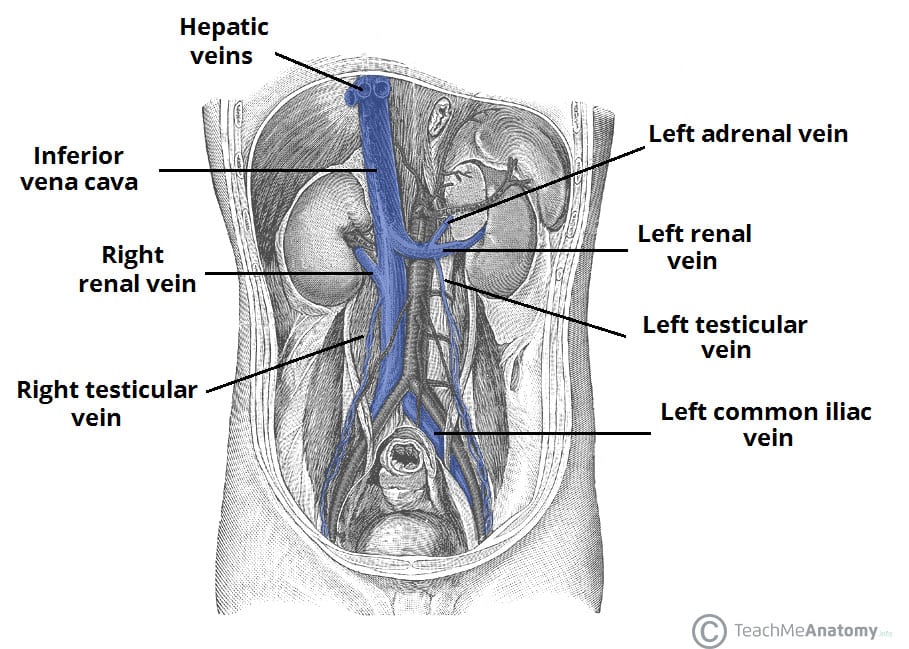
Venous Drainage of the Abdomen TeachMeAnatomy
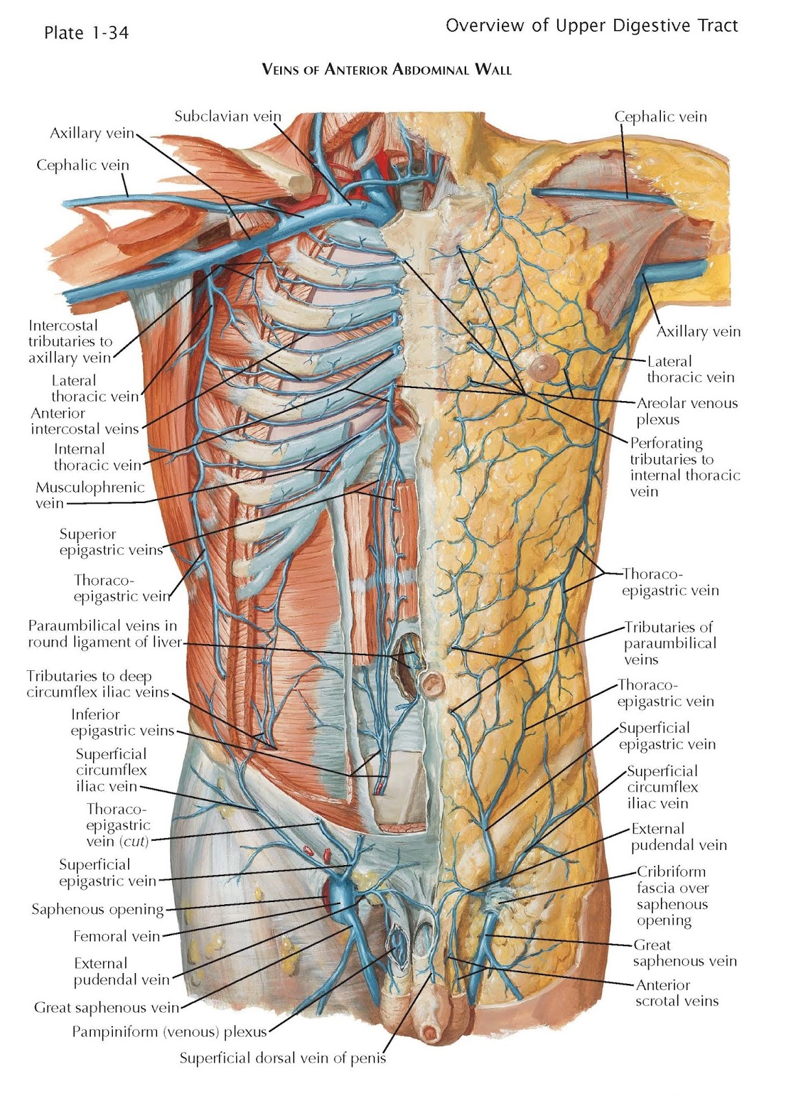
The anterior abdominal wall is drained by the superior, inferior, and superficial epigastric vessels. The arteries we've seen are accompanied by corresponding veins. Web venous.

BVS 10.2 Veins of abdomen Diagram Quizlet
Each of which provides unique microenvironmental challenges to management. The portal venous system transports venous blood from the abdominal vasculature to the liver, whilst the.
Venous Drainage of the Abdomen pediagenosis
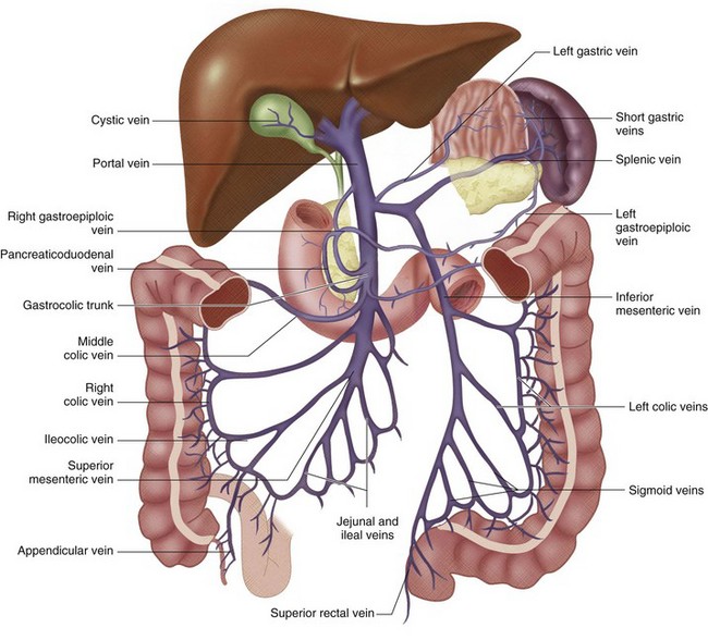
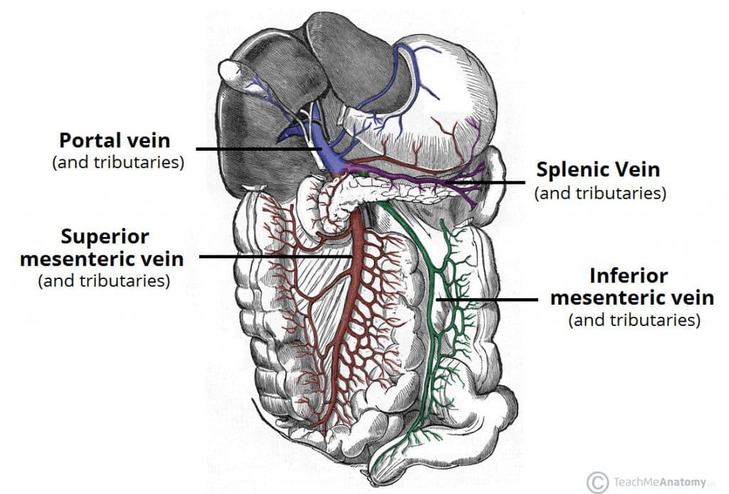
Web (2.14) now we'll move on to look at the veins that drain the gi tract, the pancreas and the spleen. Web the recognition of.
Anatomy Of Human Abdominal Vein System Digital Art by Stocktrek Images
The key difference between these two systems is the liver. The posterolateral abdominal wall is primarily drained by the tenth and eleventh posterior intercostal, subcostal,.

Abdominal Veins Labeling Diagram Quizlet
The inferior vena cava is longer than the abdominal aorta. Flow from down to up (ivc obstruction). Web this page will cover a search pattern.
Venous Anatomy of the Abdomen and Pelvis Radiology Key
In this chapter, we will review the normal anatomy and sonographic appearances of the abdominal arteries and veins. When liver disease is severe enough to.
Pictures Of Abdominal Veins
An abnormal venous pattern is a different type of abdominal marking and will be discussed separately. In this chapter, we will review the normal anatomy.
Venous Drainage of the Abdomen TeachMeAnatomy
The portal venous system is composed of the veins that drain the abdominal viscera, spleen, pancreas, and gallbladder. An abnormal venous pattern is a different.

Pictures Of Abdominal Veins
Abdominal wall and flanks for contour, masses, venous pattern and movements. Web the recognition of changes in abdominal blood flow allows accurate diagnosis of arterial.
Need To Distinguish Three Kinds Of Flow In Visible Veins.
Origin in visceral venous plexus. On grayscale (b mode) us, hyperechoic and nonshadowing speckles in the portal vein or adjacent hepatic parenchyma extending along the portal triad (preferentially peripheral and branching pattern) should. The portal system transports venous blood to the liver for processing, whilst the systemic venous system returns blood to the right atrium of the heart. Web the anterolateral abdominal wall includes the front and side walls of the abdomen.
Of Course, Recognition Of The Normal Vascular Anatomy Is Essential For The Investigation Of Any Abdominal Vascular.
This chapter will review the normal flow patterns seen in abdominal arteries and veins that will be encountered in our doppler examinations. Checking the abdominal and pelvic venous systems. Web general anatomic description. Web the recognition of changes in abdominal blood flow allows accurate diagnosis of arterial and venous abnormalities, including stenosis, occlusion, and thrombosis.
This Article Will Discuss The Anatomy Of Abdominal Arteries And Veins, As Well As Topographical Approach To The Abdominopelvic Vasculature.
The blood from the portal vein passes through the liver and finally drains into the inferior vena cava. Flow to the umbilicus (rare, in portal vein thrombosis). Nearly 80% of hepatic inflow comes from the portal vein. Abdominal wall and flanks for contour, masses, venous pattern and movements.
The Posterolateral Abdominal Wall Is Primarily Drained By The Tenth And Eleventh Posterior Intercostal, Subcostal, And Lumbar Veins.
In this chapter, we will review the normal anatomy and sonographic appearances of the abdominal arteries and veins. The venous drainage of the superficial anterolateral abdominal wall involves an elaborate subcutaneous venous plexus. The world's most advanced 3d anatomy platform. An abnormal venous pattern is a different type of abdominal marking and will be discussed separately.